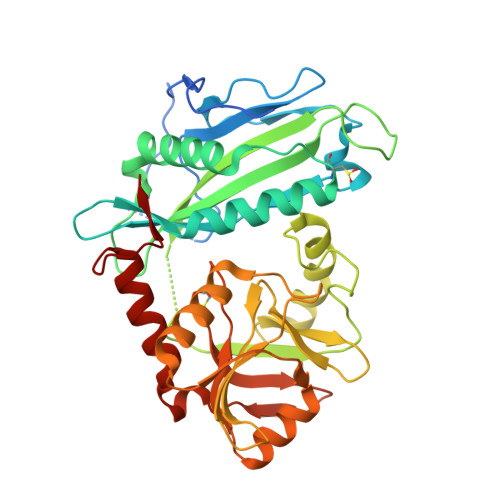
6PRX

oxidized Human Branched Chain Aminotransferase mutant C318A


Experimental Data Snapshot

  • Method: X-RAY DIFFRACTION
  • Resolution: 3.25 Å
  • R-Value Free: 0.259 
  • R-Value Work: 0.220 
  • R-Value Observed: 0.222 

wwPDB Validation   3D Report Full Report


Ligand Structure Quality Assessment 


This is version 1.2 of the entry. See complete history


Literature

Crystal structure of an oxidized mutant of human mitochondrial branched-chain aminotransferase.

Herbert, D.Gibbs, S.Riddick, A.Conway, M.Dong, M.

(2020) Acta Crystallogr F Struct Biol Commun 76: 14-19

  • DOI: https://doi.org/10.1107/S2053230X19016480
  • Primary Citation of Related Structures:  
    6PRX

  • PubMed Abstract: 

    This study presents the crystal structure of a thiol variant of the human mitochondrial branched-chain aminotransferase protein. Human branched-chain aminotransferase (hBCAT) catalyzes the transamination of the branched-chain amino acids leucine, valine and isoleucine and α-ketoglutarate to their respective α-keto acids and glutamate. hBCAT activity is regulated by a CXXC center located approximately 10 Å from the active site. This redox-active center facilitates recycling between the reduced and oxidized states, representing hBCAT in its active and inactive forms, respectively. Site-directed mutagenesis of the redox sensor (Cys315) results in a significant loss of activity, with no loss of activity reported on the mutation of the resolving cysteine (Cys318), which allows the reversible formation of a disulfide bond between Cys315 and Cys318. The crystal structure of the oxidized form of the C318A variant was used to better understand the contributions of the individual cysteines and their oxidation states. The structure reveals the modified CXXC center in a conformation similar to that in the oxidized wild type, supporting the notion that its regulatory mechanism depends on switching the Cys315 side chain between active and inactive conformations. Moreover, the structure reveals conformational differences in the N-terminal and inter-domain region that may correlate with the inactivated state of the CXXC center.


  • Organizational Affiliation

    Department of Chemistry, North Carolina Agricultural and Technical State University, USA.


Macromolecules
Find similar proteins by:  (by identity cutoff)  |  3D Structure
Entity ID: 1
MoleculeChains Sequence LengthOrganismDetailsImage
Branched-chain-amino-acid aminotransferase, mitochondrial
A, B
383Homo sapiensMutation(s): 1 
Gene Names: BCAT2BCATMBCT2ECA40
EC: 2.6.1.42
UniProt & NIH Common Fund Data Resources
Find proteins for O15382 (Homo sapiens)
Explore O15382 
Go to UniProtKB:  O15382
PHAROS:  O15382
GTEx:  ENSG00000105552 
Entity Groups  
Sequence Clusters30% Identity50% Identity70% Identity90% Identity95% Identity100% Identity
UniProt GroupO15382
Sequence Annotations
Expand
  • Reference Sequence
Small Molecules
Modified Residues  1 Unique
IDChains TypeFormula2D DiagramParent
OCS
Query on OCS
A, B
L-PEPTIDE LINKINGC3 H7 N O5 SCYS
Experimental Data & Validation

Experimental Data

  • Method: X-RAY DIFFRACTION
  • Resolution: 3.25 Å
  • R-Value Free: 0.259 
  • R-Value Work: 0.220 
  • R-Value Observed: 0.222 
  • Space Group: P 21 21 21
Unit Cell:
Length ( Å )Angle ( ˚ )
a = 69.204α = 90
b = 104.938β = 90
c = 106.294γ = 90
Software Package:
Software NamePurpose
REFMACrefinement
HKL-3000data reduction
HKL-3000data scaling
MOLREPphasing

Structure Validation

View Full Validation Report


Ligand Structure Quality Assessment 


Entry History 

Deposition Data

Revision History  (Full details and data files)

  • Version 1.0: 2020-01-22
    Type: Initial release
  • Version 1.1: 2023-10-11
    Changes: Data collection, Database references, Refinement description
  • Version 1.2: 2023-11-15
    Changes: Data collection