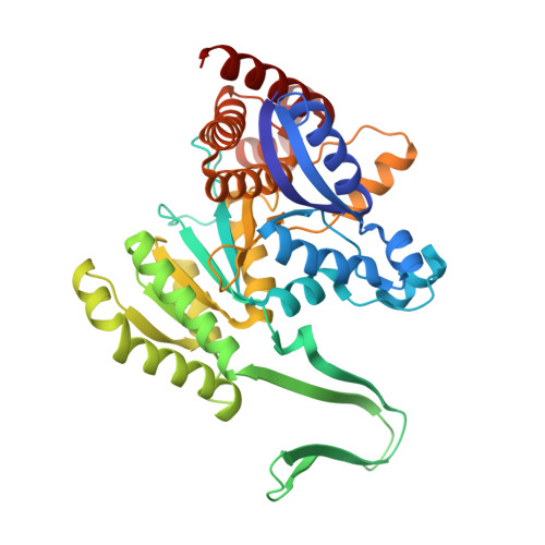
6PAY

Structure of HsICDH1:Mg(II):ICT:NADPH(50%) complex reveals structural basis for observation of half-sites reactivity


Experimental Data Snapshot

  • Method: X-RAY DIFFRACTION
  • Resolution: 2.20 Å
  • R-Value Free: 0.242 
  • R-Value Work: 0.224 
  • R-Value Observed: 0.225 

wwPDB Validation   3D Report Full Report


Ligand Structure Quality Assessment 


This is version 1.2 of the entry. See complete history


Literature

Transient-State Analysis of Human Isocitrate Dehydrogenase I: Accounting for the Interconversion of Active and Non-Active Conformational States.

Roman, J.V.Melkonian, T.R.Silvaggi, N.R.Moran, G.R.

(2019) Biochemistry 58: 5366-5380

  • DOI: https://doi.org/10.1021/acs.biochem.9b00518
  • Primary Citation of Related Structures:  
    6PAY

  • PubMed Abstract: 

    Human isocitrate dehydrogenase 1 (HsICDH1) is a cytoplasmic homodimeric Mg(II)-dependent enzyme that converts d-isocitrate (D-ICT) and NADP + to α-ketoglutarate (AKG), CO 2 , and NADPH. The active sites are formed at the subunit interface and incorporate residues from both protomers. The turnover number titrates hyperbolically from 17.5 s -1 to a minimum of 7 s -1 with an increasing enzyme concentration. As isolated, the enzyme adopts an inactive open conformation and binds NADPH tightly. The open conformation displaces three of the eight residues that bind D-ICT and Mg(II). Enzyme activation occurs with the addition of Mg(II) or D-ICT with a rate constant of 0.12 s -1 . The addition of both Mg(II) and D-ICT activates the enzyme with a rate constant of 0.6 s -1 and displaces half of the bound NADPH. This indicates that HsICDH1 may have a half-site mechanism in which the active sites alternate in catalysis. The X-ray crystal structure of the half-site activated complex reveals asymmetry in the homodimer with a single NADPH bound. The structure also indicates a pseudotetramer interface that impedes the egress of NADPH consistent with the suppression of the turnover number at high enzyme concentrations. When the half-site activated form of the enzyme is reacted with NADP + , NADPH forms with a rate constant of 204 s -1 followed by a shift in the NADPH absorption spectrum with a rate constant of 28 s -1 . These data indicate the accumulation of two intermediate states. Once D-ICT is exhausted, HsICDH1 relaxes to the inactive open state with a rate constant of ∼3 s -1 .


  • Organizational Affiliation

    Department of Chemistry and Biochemistry , Loyola University Chicago , Flanner Hall, 1068 West Sheridan Road , Chicago , Illinois 60660 , United States.


Macromolecules
Find similar proteins by:  (by identity cutoff)  |  3D Structure
Entity ID: 1
MoleculeChains Sequence LengthOrganismDetailsImage
Isocitrate dehydrogenase [NADP] cytoplasmic
A, B, C, D
414Homo sapiensMutation(s): 0 
Gene Names: IDH1PICD
EC: 1.1.1.42
UniProt & NIH Common Fund Data Resources
Find proteins for O75874 (Homo sapiens)
Explore O75874 
Go to UniProtKB:  O75874
PHAROS:  O75874
GTEx:  ENSG00000138413 
Entity Groups  
Sequence Clusters30% Identity50% Identity70% Identity90% Identity95% Identity100% Identity
UniProt GroupO75874
Sequence Annotations
Expand
  • Reference Sequence
Small Molecules
Ligands 4 Unique
IDChains Name / Formula / InChI Key2D Diagram3D Interactions
NDP
Query on NDP

Download Ideal Coordinates CCD File 
G [auth A]NADPH DIHYDRO-NICOTINAMIDE-ADENINE-DINUCLEOTIDE PHOSPHATE
C21 H30 N7 O17 P3
ACFIXJIJDZMPPO-NNYOXOHSSA-N
ICT
Query on ICT

Download Ideal Coordinates CCD File 
E [auth A],
L [auth B],
P [auth C],
R [auth D]
ISOCITRIC ACID
C6 H8 O7
ODBLHEXUDAPZAU-ZAFYKAAXSA-N
FMT
Query on FMT

Download Ideal Coordinates CCD File 
H [auth A]
I [auth A]
J [auth A]
K [auth A]
N [auth B]
H [auth A],
I [auth A],
J [auth A],
K [auth A],
N [auth B],
O [auth B],
T [auth D]
FORMIC ACID
C H2 O2
BDAGIHXWWSANSR-UHFFFAOYSA-N
MG
Query on MG

Download Ideal Coordinates CCD File 
F [auth A],
M [auth B],
Q [auth C],
S [auth D]
MAGNESIUM ION
Mg
JLVVSXFLKOJNIY-UHFFFAOYSA-N
Binding Affinity Annotations 
IDSourceBinding Affinity
ICT Binding MOAD:  6PAY Kd: 5.10e+5 (nM) from 1 assay(s)
Experimental Data & Validation

Experimental Data

  • Method: X-RAY DIFFRACTION
  • Resolution: 2.20 Å
  • R-Value Free: 0.242 
  • R-Value Work: 0.224 
  • R-Value Observed: 0.225 
  • Space Group: P 1 21 1
Unit Cell:
Length ( Å )Angle ( ˚ )
a = 94.6α = 90
b = 74.6β = 95.2
c = 142.9γ = 90
Software Package:
Software NamePurpose
PHENIXrefinement
HKL-2000data reduction
HKL-2000data scaling
PHASERphasing

Structure Validation

View Full Validation Report


Ligand Structure Quality Assessment 


Entry History 

Deposition Data

Revision History  (Full details and data files)

  • Version 1.0: 2019-09-18
    Type: Initial release
  • Version 1.1: 2020-01-08
    Changes: Database references
  • Version 1.2: 2023-10-11
    Changes: Data collection, Database references, Derived calculations, Refinement description