6OAM

Crystal Structure of ChlaDUB2 DUB domain


Experimental Data Snapshot

  • Method: X-RAY DIFFRACTION
  • Resolution: 2.50 Å
  • R-Value Free: 0.306 
  • R-Value Work: 0.283 
  • R-Value Observed: 0.284 

wwPDB Validation   3D Report Full Report

Currently 6OAM does not have a validation slider image.


This is version 1.2 of the entry. See complete history


Literature

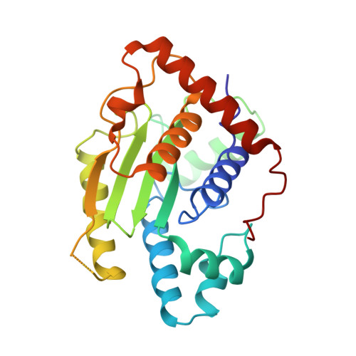
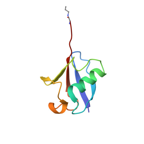
The Two Deubiquitinating Enzymes fromChlamydia trachomatisHave Distinct Ubiquitin Recognition Properties.

Hausman, J.M.Kenny, S.Iyer, S.Babar, A.Qiu, J.Fu, J.Luo, Z.Q.Das, C.

(2020) Biochemistry 59: 1604-1617

  • DOI: https://doi.org/10.1021/acs.biochem.9b01107
  • Primary Citation of Related Structures:  
    6MRN, 6OAM

  • PubMed Abstract: 

    Chlamydia trachomatis is the cause of several diseases such as sexually transmitted urogenital disease and ocular trachoma. The pathogen contains a small genome yet, upon infection, expresses two enzymes with deubiquitinating activity, termed ChlaDUB1 and ChlaDUB2, presumed to have redundant deubiquitinase (DUB) function because of the similarity of the primary structure of their catalytic domain. Previous studies have led to structural characterization of the enzymatic properties of ChlaDUB1; however, ChlaDUB2 has yet to be investigated thoroughly. In this study, we investigated the deubiquitinase properties of ChlaDUB2 and compared them to those of ChlaDUB1. This revealed a distinct difference in hydrolytic activity with regard to di- and polyubiquitin chains while showing similar ability to cleave a monoubiquitin-based substrate, ubiquitin aminomethylcoumarin (Ub-AMC). ChlaDUB2 was unable to cleave a diubiquitin substrate efficiently, whereas ChlaDUB1 could rapidly hydrolyze this substrate like a prototypical prokaryotic DUB, SdeA. With polyubiquitinated green fluorescent protein substrate (GFP-Ub n ), whereas ChlaDUB1 efficiently disassembled the polyubiquitin chains into the monoubiquitin product, the deubiquitination activity of ChlaDUB2, while showing depletion of the substrate, did not produce appreciable levels of the monoubiquitin product. We report the structures of a catalytic construct of ChlaDUB2 and its complex with ubiquitin propargyl amide. These structures revealed differences in residues involved in substrate recognition between the two Chlamydia DUBs. On the basis of the structures, we conclude that the distal ubiquitin binding is equivalent between the two DUBs, consistent with the Ub-AMC activity result. Therefore, the difference in activity with longer ubiquitinated substrates may be due to the differential recognition of these substrates involving additional ubiquitin binding sites.


  • Organizational Affiliation

    Department of Chemistry, Purdue University, 560 Oval Drive, West Lafayette, Indiana 47907, United States.


Macromolecules
Find similar proteins by:  (by identity cutoff)  |  3D Structure
Entity ID: 1
MoleculeChains Sequence LengthOrganismDetailsImage
Deubiquitinase and deneddylase Dub2A,
C [auth B]
251Chlamydia trachomatis L2/434/BuMutation(s): 0 
Gene Names: cdu2CTL0246
EC: 3.4.22
UniProt
Find proteins for B0B999 (Chlamydia trachomatis serovar L2 (strain 434/Bu / ATCC VR-902B))
Explore B0B999 
Go to UniProtKB:  B0B999
Entity Groups  
Sequence Clusters30% Identity50% Identity70% Identity90% Identity95% Identity100% Identity
UniProt GroupB0B999
Sequence Annotations
Expand
  • Reference Sequence
Find similar proteins by:  (by identity cutoff)  |  3D Structure
Entity ID: 2
MoleculeChains Sequence LengthOrganismDetailsImage
UbiquitinB [auth D],
D [auth C]
76Homo sapiensMutation(s): 1 
Gene Names: UBB
UniProt & NIH Common Fund Data Resources
Find proteins for P0CG47 (Homo sapiens)
Explore P0CG47 
Go to UniProtKB:  P0CG47
PHAROS:  P0CG47
GTEx:  ENSG00000170315 
Entity Groups  
Sequence Clusters30% Identity50% Identity70% Identity90% Identity95% Identity100% Identity
UniProt GroupP0CG47
Sequence Annotations
Expand
  • Reference Sequence
Experimental Data & Validation

Experimental Data

  • Method: X-RAY DIFFRACTION
  • Resolution: 2.50 Å
  • R-Value Free: 0.306 
  • R-Value Work: 0.283 
  • R-Value Observed: 0.284 
  • Space Group: P 43
Unit Cell:
Length ( Å )Angle ( ˚ )
a = 108.692α = 90
b = 108.692β = 90
c = 62.278γ = 90
Software Package:
Software NamePurpose
PHENIXrefinement
HKL-2000data reduction
SCALEPACKdata scaling
PHASERphasing

Structure Validation

View Full Validation Report

Currently 6OAM does not have a validation slider image.


Entry History & Funding Information

Deposition Data


Funding OrganizationLocationGrant Number
National Institutes of Health/National Human Genome Research Institute (NIH/NHGRI)United States2R01GM103401

Revision History  (Full details and data files)

  • Version 1.0: 2020-04-22
    Type: Initial release
  • Version 1.1: 2020-05-13
    Changes: Database references
  • Version 1.2: 2023-10-11
    Changes: Advisory, Data collection, Database references, Refinement description