6N3C

SegB, conformation of TDP-43 low complexity domain segment A


Experimental Data Snapshot

  • Method: ELECTRON MICROSCOPY
  • Resolution: 3.30 Å
  • Aggregation State: FILAMENT 
  • Reconstruction Method: HELICAL 

wwPDB Validation   3D Report Full Report


This is version 1.4 of the entry. See complete history


Literature

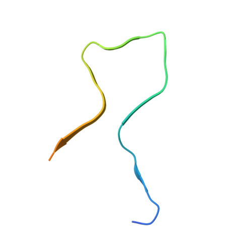
Cryo-EM structures of four polymorphic TDP-43 amyloid cores.

Cao, Q.Boyer, D.R.Sawaya, M.R.Ge, P.Eisenberg, D.S.

(2019) Nat Struct Mol Biol 26: 619-627

  • DOI: https://doi.org/10.1038/s41594-019-0248-4
  • Primary Citation of Related Structures:  
    6N37, 6N3A, 6N3B, 6N3C

  • PubMed Abstract: 

    The DNA and RNA processing protein TDP-43 undergoes both functional and pathogenic aggregation. Functional TDP-43 aggregates form reversible, transient species such as nuclear bodies, stress granules, and myo-granules. Pathogenic, irreversible TDP-43 aggregates form in amyotrophic lateral sclerosis and other neurodegenerative conditions. Here we find the features of TDP-43 fibrils that confer both reversibility and irreversibility by determining structures of two segments reported to be the pathogenic cores of human TDP-43 aggregation: SegA (residues 311-360), which forms three polymorphs, all with dagger-shaped folds; and SegB A315E (residues 286-331 containing the amyotrophic lateral sclerosis hereditary mutation A315E), which forms R-shaped folds. Energetic analysis suggests that the dagger-shaped polymorphs represent irreversible fibril structures, whereas the SegB polymorph may participate in both reversible and irreversible fibrils. Our structures reveal the polymorphic nature of TDP-43 and suggest how the A315E mutation converts the R-shaped polymorph to an irreversible form that enhances pathology.


  • Organizational Affiliation

    Department of Chemistry and Biochemistry and Biological Chemistry, UCLA-DOE Institute, Molecular Biology Institute, and Howard Hughes Medical Institute, UCLA, Los Angeles, CA, USA.


Macromolecules
Find similar proteins by:  (by identity cutoff)  |  3D Structure
Entity ID: 1
MoleculeChains Sequence LengthOrganismDetailsImage
TAR DNA-binding protein 43
A, B, C, D, E
A, B, C, D, E, F, G, H, I, J, K, L, M, N, O, P, Q, R, S, T
46Homo sapiensMutation(s): 0 
Gene Names: TARDBPTDP43
UniProt & NIH Common Fund Data Resources
Find proteins for Q13148 (Homo sapiens)
Explore Q13148 
Go to UniProtKB:  Q13148
PHAROS:  Q13148
GTEx:  ENSG00000120948 
Entity Groups  
Sequence Clusters30% Identity50% Identity70% Identity90% Identity95% Identity100% Identity
UniProt GroupQ13148
Sequence Annotations
Expand
  • Reference Sequence
Experimental Data & Validation

Experimental Data

  • Method: ELECTRON MICROSCOPY
  • Resolution: 3.30 Å
  • Aggregation State: FILAMENT 
  • Reconstruction Method: HELICAL 
EM Software:
TaskSoftware PackageVersion
MODEL REFINEMENTPHENIXreal_space_refine
RECONSTRUCTIONRELION2

Structure Validation

View Full Validation Report


Entry History & Funding Information

Deposition Data


Funding OrganizationLocationGrant Number
National Institutes of Health/National Institute on Aging (NIH/NIA)United StatesAG0543022

Revision History  (Full details and data files)

  • Version 1.0: 2019-06-26
    Type: Initial release
  • Version 1.1: 2019-07-10
    Changes: Data collection, Database references
  • Version 1.2: 2019-07-17
    Changes: Data collection, Database references
  • Version 1.3: 2019-12-18
    Changes: Author supporting evidence
  • Version 1.4: 2024-03-20
    Changes: Data collection, Database references