6L1I

Crystal Structure Of of PHF20L1 Tudor1 Y24W/Y29W mutant


Experimental Data Snapshot

  • Method: X-RAY DIFFRACTION
  • Resolution: 1.85 Å
  • R-Value Free: 0.208 
  • R-Value Work: 0.160 
  • R-Value Observed: 0.164 

wwPDB Validation   3D Report Full Report


This is version 1.2 of the entry. See complete history


Literature

Conformational Selection in Ligand Recognition by the First Tudor Domain of PHF20L1.

Lv, M.Gao, J.Li, M.Ma, R.Li, F.Liu, Y.Liu, M.Zhang, J.Yao, X.Wu, J.Shi, Y.Tang, Y.Pan, Y.Zhang, Z.Ruan, K.

(2020) J Phys Chem Lett 11: 7932-7938

  • DOI: https://doi.org/10.1021/acs.jpclett.0c02039
  • Primary Citation of Related Structures:  
    6L0X, 6L10, 6L1C, 6L1F, 6L1I, 6L1P

  • PubMed Abstract: 

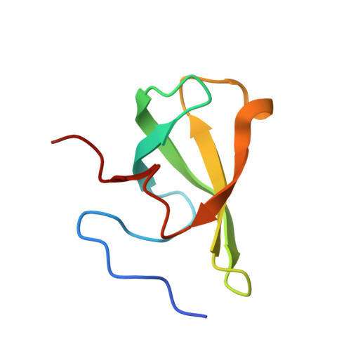
    The first Tudor domain (Tudor1) of PHF20L1 recognizes (non)histone methylation to play versatile roles. However, the underlying ligand-recognition mechanism remains unknown as a closed state revealed in the free-form structure. NMR relaxation dispersion and molecular dynamics simulations suggest a pre-existing low-population conformation with a remarkable rearrangement of aromatic cage residues of PHF20L1 Tudor1. Such an open-form conformation is utilized to recognize lysine 142 methylated DNMT1, a cosolvent, and an NMR fragment screening hit, as revealed by the complex crystal structures. Intriguingly, the ligand binding capacity was enhanced by mutation that tunes up the open-state population only. The recognition of DNMT1 by PHF20L1 was further validated in cancer cells. This conformational selection mechanism will enable the discovery of small molecule inhibitors against the seemingly "undruggable" PHF20L1 Tudor1.


  • Organizational Affiliation

    Ministry of Education Key Laboratory for Membrane-less Organelles & Cellular Dynamics, Hefei National Laboratory for Physical Sciences at the Microscale, the First Affiliated Hospital & School of Life Sciences, University of Science and Technology of China, Hefei, Anhui 230027, P.R. China.


Macromolecules
Find similar proteins by:  (by identity cutoff)  |  3D Structure
Entity ID: 1
MoleculeChains Sequence LengthOrganismDetailsImage
PHD finger protein 20-like protein 1A [auth B]70Homo sapiensMutation(s): 2 
Gene Names: PHF20L1CGI-72
UniProt & NIH Common Fund Data Resources
Find proteins for A8MW92 (Homo sapiens)
Explore A8MW92 
Go to UniProtKB:  A8MW92
PHAROS:  A8MW92
GTEx:  ENSG00000129292 
Entity Groups  
Sequence Clusters30% Identity50% Identity70% Identity90% Identity95% Identity100% Identity
UniProt GroupA8MW92
Sequence Annotations
Expand
  • Reference Sequence
Small Molecules
Experimental Data & Validation

Experimental Data

  • Method: X-RAY DIFFRACTION
  • Resolution: 1.85 Å
  • R-Value Free: 0.208 
  • R-Value Work: 0.160 
  • R-Value Observed: 0.164 
  • Space Group: P 65
Unit Cell:
Length ( Å )Angle ( ˚ )
a = 63.124α = 90
b = 63.124β = 90
c = 26.98γ = 120
Software Package:
Software NamePurpose
PHENIXrefinement
SCALAdata scaling
PDB_EXTRACTdata extraction
HKL-2000data reduction
PHASERphasing

Structure Validation

View Full Validation Report


Entry History 

Deposition Data

Revision History  (Full details and data files)

  • Version 1.0: 2020-09-23
    Type: Initial release
  • Version 1.1: 2020-09-30
    Changes: Database references
  • Version 1.2: 2023-11-22
    Changes: Data collection, Database references, Refinement description