Covalent Aurora A regulation by the metabolic integrator coenzyme A.
Tsuchiya, Y., Byrne, D.P., Burgess, S.G., Bormann, J., Bakovic, J., Huang, Y., Zhyvoloup, A., Yu, B.Y.K., Peak-Chew, S., Tran, T., Bellany, F., Tabor, A.B., Chan, A.E., Guruprasad, L., Garifulin, O., Filonenko, V., Vonderach, M., Ferries, S., Eyers, C.E., Carroll, J., Skehel, M., Bayliss, R., Eyers, P.A., Gout, I.(2019) Redox Biol 28: 101318-101318
- PubMed: 31546169
- DOI: https://doi.org/10.1016/j.redox.2019.101318
- Primary Citation of Related Structures:
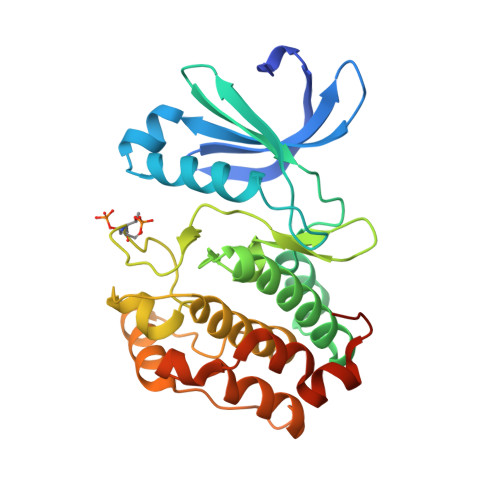
6I2U - PubMed Abstract:
Aurora A kinase is a master mitotic regulator whose functions are controlled by several regulatory interactions and post-translational modifications. It is frequently dysregulated in cancer, making Aurora A inhibition a very attractive antitumor target. However, recently uncovered links between Aurora A, cellular metabolism and redox regulation are not well understood. In this study, we report a novel mechanism of Aurora A regulation in the cellular response to oxidative stress through CoAlation. A combination of biochemical, biophysical, crystallographic and cell biology approaches revealed a new and, to our knowledge, unique mode of Aurora A inhibition by CoA, involving selective binding of the ADP moiety of CoA to the ATP binding pocket and covalent modification of Cys290 in the activation loop by the thiol group of the pantetheine tail. We provide evidence that covalent CoA modification (CoAlation) of Aurora A is specific, and that it can be induced by oxidative stress in human cells. Oxidising agents, such as diamide, hydrogen peroxide and menadione were found to induce Thr 288 phosphorylation and DTT-dependent dimerization of Aurora A. Moreover, microinjection of CoA into fertilized mouse embryos disrupts bipolar spindle formation and the alignment of chromosomes, consistent with Aurora A inhibition. Altogether, our data reveal CoA as a new, rather selective, inhibitor of Aurora A, which locks this kinase in an inactive state via a "dual anchor" mechanism of inhibition that might also operate in cellular response to oxidative stress. Finally and most importantly, we believe that these novel findings provide a new rationale for developing effective and irreversible inhibitors of Aurora A, and perhaps other protein kinases containing appropriately conserved Cys residues.
Organizational Affiliation:
Department of Structural and Molecular Biology, University College London, London, WC1E 6BT, UK.