6HPY

Crystal structure of ENL (MLLT1) in complex with compound 12


Experimental Data Snapshot

  • Method: X-RAY DIFFRACTION
  • Resolution: 2.00 Å
  • R-Value Free: 0.260 
  • R-Value Work: 0.217 
  • R-Value Observed: 0.219 

wwPDB Validation   3D Report Full Report


Ligand Structure Quality Assessment 


This is version 1.3 of the entry. See complete history


Literature

Structure-Based Approach toward Identification of Inhibitory Fragments for Eleven-Nineteen-Leukemia Protein (ENL).

Heidenreich, D.Moustakim, M.Schmidt, J.Merk, D.Brennan, P.E.Fedorov, O.Chaikuad, A.Knapp, S.

(2018) J Med Chem 61: 10929-10934

  • DOI: https://doi.org/10.1021/acs.jmedchem.8b01457
  • Primary Citation of Related Structures:  
    6HPW, 6HPX, 6HPY, 6HPZ, 6HQ0

  • PubMed Abstract: 

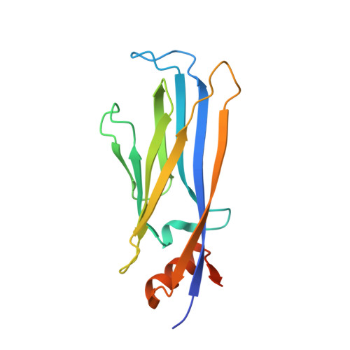
    Lysine acetylation is an epigenetic mark that is principally recognized by bromodomains, and recently structurally diverse YEATS domains also emerged as readers of lysine acetyl/acylations. Here we present a crystallography-based strategy and the discovery of fragments binding to the ENL YEATS domain, a potential drug target. Crystal structures combined with synthetic efforts led to the identification of a submicromolar binder, providing first starting points for the development of chemical probes for this reader domain family.


  • Organizational Affiliation

    Institute of Pharmaceutical Chemistry , Goethe-University Frankfurt , 60438 Frankfurt , Germany.


Macromolecules
Find similar proteins by:  (by identity cutoff)  |  3D Structure
Entity ID: 1
MoleculeChains Sequence LengthOrganismDetailsImage
Protein ENL155Homo sapiensMutation(s): 0 
Gene Names: MLLT1ENLLTG19YEATS1
UniProt & NIH Common Fund Data Resources
Find proteins for Q03111 (Homo sapiens)
Explore Q03111 
Go to UniProtKB:  Q03111
PHAROS:  Q03111
GTEx:  ENSG00000130382 
Entity Groups  
Sequence Clusters30% Identity50% Identity70% Identity90% Identity95% Identity100% Identity
UniProt GroupQ03111
Sequence Annotations
Expand
  • Reference Sequence
Experimental Data & Validation

Experimental Data

  • Method: X-RAY DIFFRACTION
  • Resolution: 2.00 Å
  • R-Value Free: 0.260 
  • R-Value Work: 0.217 
  • R-Value Observed: 0.219 
  • Space Group: P 43 21 2
Unit Cell:
Length ( Å )Angle ( ˚ )
a = 49.039α = 90
b = 49.039β = 90
c = 132.729γ = 90
Software Package:
Software NamePurpose
REFMACrefinement
iMOSFLMdata reduction
Aimlessdata scaling
PHASERphasing

Structure Validation

View Full Validation Report


Ligand Structure Quality Assessment 


Entry History 

Deposition Data

Revision History  (Full details and data files)

  • Version 1.0: 2018-11-28
    Type: Initial release
  • Version 1.1: 2018-12-05
    Changes: Data collection, Database references
  • Version 1.2: 2019-04-24
    Changes: Data collection, Database references
  • Version 1.3: 2024-01-24
    Changes: Data collection, Database references, Refinement description