6GZY

HOIP-fragment5 complex


Experimental Data Snapshot

  • Method: X-RAY DIFFRACTION
  • Resolution: 2.15 Å
  • R-Value Free: 0.246 
  • R-Value Work: 0.199 
  • R-Value Observed: 0.201 

wwPDB Validation   3D Report Full Report


Ligand Structure Quality Assessment 


This is version 1.2 of the entry. See complete history


Literature

Fragment-Based Covalent Ligand Screening Enables Rapid Discovery of Inhibitors for the RBR E3 Ubiquitin Ligase HOIP.

Johansson, H.Isabella Tsai, Y.C.Fantom, K.Chung, C.W.Kumper, S.Martino, L.Thomas, D.A.Eberl, H.C.Muelbaier, M.House, D.Rittinger, K.

(2019) J Am Chem Soc 141: 2703-2712

  • DOI: https://doi.org/10.1021/jacs.8b13193
  • Primary Citation of Related Structures:  
    6GZY

  • PubMed Abstract: 

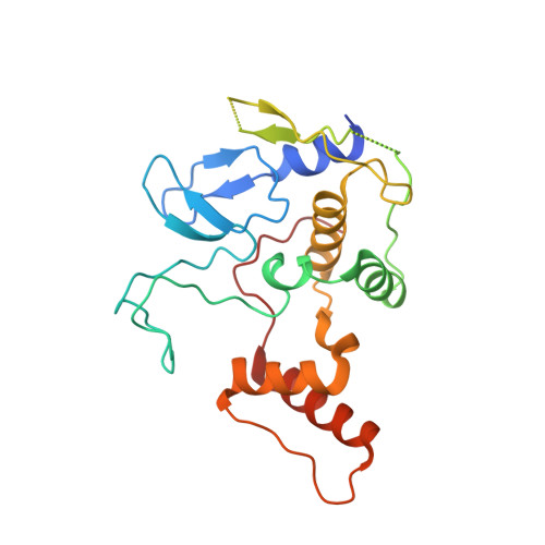
    Modification of proteins with polyubiquitin chains is a key regulatory mechanism to control cellular behavior and alterations in the ubiquitin system are linked to many diseases. Linear (M1-linked) polyubiquitin chains play pivotal roles in several cellular signaling pathways mediating immune and inflammatory responses and apoptotic cell death. These chains are formed by the linear ubiquitin chain assembly complex (LUBAC), a multiprotein E3 ligase that consists of 3 subunits, HOIP, HOIL-1L, and SHARPIN. Herein, we describe the discovery of inhibitors targeting the active site cysteine of the catalytic subunit HOIP using fragment-based covalent ligand screening. We report the synthesis of a diverse library of electrophilic fragments and demonstrate an integrated use of protein LC-MS, biochemical ubiquitination assays, chemical synthesis, and protein crystallography to enable the first structure-based development of covalent inhibitors for an RBR E3 ligase. Furthermore, using cell-based assays and chemoproteomics, we demonstrate that these compounds effectively penetrate mammalian cells to label and inhibit HOIP and NF-κB activation, making them suitable hits for the development of selective probes to study LUBAC biology. Our results illustrate the power of fragment-based covalent ligand screening to discover lead compounds for challenging targets, which holds promise to be a general approach for the development of cell-permeable inhibitors of thioester-forming E3 ubiquitin ligases.


  • Organizational Affiliation

    Crick-GSK Biomedical LinkLabs , GlaxoSmithKline , Gunnels Wood Road , Stevenage SG1 2NY , United Kingdom.


Macromolecules
Find similar proteins by:  (by identity cutoff)  |  3D Structure
Entity ID: 1
MoleculeChains Sequence LengthOrganismDetailsImage
E3 ubiquitin-protein ligase RNF31
A, B
223Homo sapiensMutation(s): 0 
Gene Names: RNF31ZIBRA
EC: 2.3.2.27
UniProt & NIH Common Fund Data Resources
Find proteins for Q96EP0 (Homo sapiens)
Explore Q96EP0 
Go to UniProtKB:  Q96EP0
PHAROS:  Q96EP0
GTEx:  ENSG00000092098 
Entity Groups  
Sequence Clusters30% Identity50% Identity70% Identity90% Identity95% Identity100% Identity
UniProt GroupQ96EP0
Sequence Annotations
Expand
  • Reference Sequence
Small Molecules
Ligands 6 Unique
IDChains Name / Formula / InChI Key2D Diagram3D Interactions
FHH
Query on FHH

Download Ideal Coordinates CCD File 
C [auth A],
P [auth B]
methyl 4-[(2-oxidanylidene-1,5,6,7-tetrahydrocyclopenta[b]pyridin-3-yl)carbonylamino]butanoate
C14 H18 N2 O4
LFOKUSURFOQCKJ-UHFFFAOYSA-N
PGE
Query on PGE

Download Ideal Coordinates CCD File 
N [auth A]TRIETHYLENE GLYCOL
C6 H14 O4
ZIBGPFATKBEMQZ-UHFFFAOYSA-N
SO4
Query on SO4

Download Ideal Coordinates CCD File 
H [auth A]
I [auth A]
J [auth A]
K [auth A]
U [auth B]
H [auth A],
I [auth A],
J [auth A],
K [auth A],
U [auth B],
V [auth B],
W [auth B]
SULFATE ION
O4 S
QAOWNCQODCNURD-UHFFFAOYSA-L
ZN
Query on ZN

Download Ideal Coordinates CCD File 
D [auth A]
E [auth A]
F [auth A]
G [auth A]
Q [auth B]
D [auth A],
E [auth A],
F [auth A],
G [auth A],
Q [auth B],
R [auth B],
S [auth B],
T [auth B]
ZINC ION
Zn
PTFCDOFLOPIGGS-UHFFFAOYSA-N
EDO
Query on EDO

Download Ideal Coordinates CCD File 
L [auth A],
M [auth A],
X [auth B],
Y [auth B]
1,2-ETHANEDIOL
C2 H6 O2
LYCAIKOWRPUZTN-UHFFFAOYSA-N
NA
Query on NA

Download Ideal Coordinates CCD File 
O [auth A],
Z [auth B]
SODIUM ION
Na
FKNQFGJONOIPTF-UHFFFAOYSA-N
Experimental Data & Validation

Experimental Data

  • Method: X-RAY DIFFRACTION
  • Resolution: 2.15 Å
  • R-Value Free: 0.246 
  • R-Value Work: 0.199 
  • R-Value Observed: 0.201 
  • Space Group: P 21 21 2
Unit Cell:
Length ( Å )Angle ( ˚ )
a = 111.202α = 90
b = 117.249β = 90
c = 37.94γ = 90
Software Package:
Software NamePurpose
PHENIXrefinement
XDSdata reduction
autoPROCdata scaling
Aimlessdata scaling
pointlessdata scaling
PHASERphasing

Structure Validation

View Full Validation Report


Ligand Structure Quality Assessment 


Entry History & Funding Information

Deposition Data


Funding OrganizationLocationGrant Number
United KingdomFC001142

Revision History  (Full details and data files)

  • Version 1.0: 2019-01-30
    Type: Initial release
  • Version 1.1: 2019-02-20
    Changes: Data collection, Database references
  • Version 1.2: 2024-01-17
    Changes: Advisory, Data collection, Database references, Derived calculations, Refinement description