6FVE

Macrophage Migration Inhibitory Factor (MIF) with Covalently Bound FITC


Experimental Data Snapshot

  • Method: X-RAY DIFFRACTION
  • Resolution: 1.41 Å
  • R-Value Free: 0.189 
  • R-Value Work: 0.134 
  • R-Value Observed: 0.136 

wwPDB Validation   3D Report Full Report


Ligand Structure Quality Assessment 


This is version 1.2 of the entry. See complete history


Literature

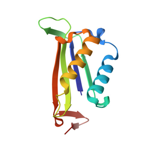
Structural Study of the Complex Formed by Ceruloplasmin and Macrophage Migration Inhibitory Factor.

Sokolov, A.V.Dadinova, L.A.Petoukhov, M.V.Bourenkov, G.Dubova, K.M.Amarantov, S.V.Volkov, V.V.Kostevich, V.A.Gorbunov, N.P.Grudinina, N.A.Vasilyev, V.B.Samygina, V.R.

(2018) Biochemistry (Mosc) 83: 701-707

  • DOI: https://doi.org/10.1134/S000629791806007X
  • Primary Citation of Related Structures:  
    6FVE, 6FVH

  • PubMed Abstract: 

    Macrophage migration inhibitory factor (MIF) is a key proinflammatory cytokine. Inhibitors of tautomerase activity of MIF are perspective antiinflammatory compounds. Ceruloplasmin, the copper-containing ferroxidase of blood plasma, is a noncompetitive inhibitor of tautomerase activity of MIF in the reaction with p-hydroxyphenylpyruvate. Small-angle X-ray scattering established a model of the complex formed by MIF and ceruloplasmin. Crystallographic analysis of MIF with a modified active site supports the model. The stoichiometry of 3 CP/MIF trimer complex was established using gel filtration. Conformity of novel data concerning the interaction regions in the studied proteins with previous biochemical data is discussed.


  • Organizational Affiliation

    Institute of Experimental Medicine, St. Petersburg, 197376, Russia.


Macromolecules
Find similar proteins by:  (by identity cutoff)  |  3D Structure
Entity ID: 1
MoleculeChains Sequence LengthOrganismDetailsImage
Macrophage migration inhibitory factorA,
B [auth C],
C [auth B]
114Homo sapiensMutation(s): 0 
Gene Names: MIFGLIFMMIF
EC: 5.3.2.1 (PDB Primary Data), 5.3.3.12 (PDB Primary Data)
UniProt & NIH Common Fund Data Resources
Find proteins for P14174 (Homo sapiens)
Explore P14174 
Go to UniProtKB:  P14174
PHAROS:  P14174
GTEx:  ENSG00000240972 
Entity Groups  
Sequence Clusters30% Identity50% Identity70% Identity90% Identity95% Identity100% Identity
UniProt GroupP14174
Sequence Annotations
Expand
  • Reference Sequence
Experimental Data & Validation

Experimental Data

  • Method: X-RAY DIFFRACTION
  • Resolution: 1.41 Å
  • R-Value Free: 0.189 
  • R-Value Work: 0.134 
  • R-Value Observed: 0.136 
  • Space Group: P 21 21 21
Unit Cell:
Length ( Å )Angle ( ˚ )
a = 46.783α = 90
b = 70.644β = 90
c = 89.8γ = 90
Software Package:
Software NamePurpose
REFMACrefinement
XDSdata reduction
Aimlessdata scaling
MOLREPphasing

Structure Validation

View Full Validation Report


Ligand Structure Quality Assessment 


Entry History & Funding Information

Deposition Data


Funding OrganizationLocationGrant Number
RFBRRussian Federation15-54-74006

Revision History  (Full details and data files)

  • Version 1.0: 2018-06-06
    Type: Initial release
  • Version 1.1: 2018-09-19
    Changes: Data collection, Database references
  • Version 1.2: 2024-01-17
    Changes: Data collection, Database references, Refinement description