6FAW

Crystal structure of C-terminal modified Tau peptide-hybrid 4.2c-I with 14-3-3sigma


Experimental Data Snapshot

  • Method: X-RAY DIFFRACTION
  • Resolution: 1.40 Å
  • R-Value Free: 0.171 
  • R-Value Work: 0.143 
  • R-Value Observed: 0.144 

wwPDB Validation   3D Report Full Report


Ligand Structure Quality Assessment 


This is version 1.3 of the entry. See complete history


Literature

Inhibition of 14-3-3/Tau by Hybrid Small-Molecule Peptides Operating via Two Different Binding Modes.

Andrei, S.A.Meijer, F.A.Neves, J.F.Brunsveld, L.Landrieu, I.Ottmann, C.Milroy, L.G.

(2018) ACS Chem Neurosci 9: 2639-2654

  • DOI: https://doi.org/10.1021/acschemneuro.8b00118
  • Primary Citation of Related Structures:  
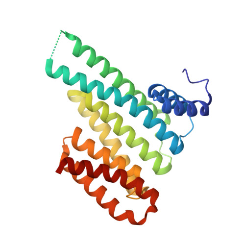
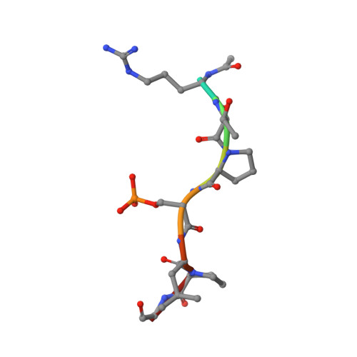
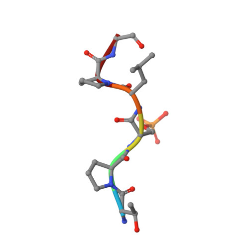
    6FAU, 6FAV, 6FAW, 6FBW, 6FBY, 6FI4, 6FI5

  • PubMed Abstract: 

    Current molecular hypotheses have not yet delivered marketable treatments for Alzheimer's disease (AD), arguably due to a lack of understanding of AD biology and an overreliance on conventional drug modalities. Protein-protein interactions (PPIs) are emerging drug targets, which show promise for the treatment of, e.g., cancer, but are still underexploited for treating neurodegenerative diseases. 14-3-3 binding to phosphorylated Tau is a promising PPI drug target based on its reported destabilizing effect on microtubules, leading to enhanced neurofibrillary tangle formation as a potential cause of AD-related neurodegeneration. Inhibition of 14-3-3/Tau may therefore be neuroprotective. Previously, we reported the structure-guided development of modified peptide inhibitors of 14-3-3/Tau. Here, we report further efforts to optimize the binding mode and activity of our modified Tau peptides through a combination of chemical synthesis, biochemical assays, and X-ray crystallography. Most notably, we were able to characterize two different high-affinity binding modes, both of which inhibited 14-3-3-binding to full-length PKA-phosphorylated Tau protein in vitro as measured by NMR spectroscopy. Our findings, besides producing useful tool inhibitor compounds for studying 14-3-3/Tau, have enhanced our understanding of the molecular parameters for inhibiting 14-3-3/Tau, which are important milestones toward the establishment of our 14-3-3 PPI hypothesis.


  • Organizational Affiliation

    Laboratory of Chemical Biology, Department of Biomedical Engineering and Institute for Complex Molecular Systems , Technische Universiteit Eindhoven , Den Dolech 2 , 5612 AZ Eindhoven , The Netherlands.


Macromolecules
Find similar proteins by:  (by identity cutoff)  |  3D Structure
Entity ID: 1
MoleculeChains Sequence LengthOrganismDetailsImage
14-3-3 protein sigma
A, C
236Homo sapiensMutation(s): 0 
Gene Names: SFNHME1
UniProt & NIH Common Fund Data Resources
Find proteins for P31947 (Homo sapiens)
Explore P31947 
Go to UniProtKB:  P31947
PHAROS:  P31947
GTEx:  ENSG00000175793 
Entity Groups  
Sequence Clusters30% Identity50% Identity70% Identity90% Identity95% Identity100% Identity
UniProt GroupP31947
Sequence Annotations
Expand
  • Reference Sequence

Find similar proteins by:  Sequence   |   3D Structure  

Entity ID: 2
MoleculeChains Sequence LengthOrganismDetailsImage
ACE-ARG-THR-PRO-SEP-LEU-PRO-GLY8Homo sapiensMutation(s): 0 
UniProt & NIH Common Fund Data Resources
Find proteins for P10636 (Homo sapiens)
Explore P10636 
Go to UniProtKB:  P10636
PHAROS:  P10636
GTEx:  ENSG00000186868 
Entity Groups  
UniProt GroupP10636
Sequence Annotations
Expand
  • Reference Sequence

Find similar proteins by:  Sequence   |   3D Structure  

Entity ID: 3
MoleculeChains Sequence LengthOrganismDetailsImage
THR-PRO-SEP-LEU-PRO-GLY6Homo sapiensMutation(s): 0 
UniProt & NIH Common Fund Data Resources
Find proteins for P10636 (Homo sapiens)
Explore P10636 
Go to UniProtKB:  P10636
PHAROS:  P10636
GTEx:  ENSG00000186868 
Entity Groups  
UniProt GroupP10636
Sequence Annotations
Expand
  • Reference Sequence
Small Molecules
Ligands 4 Unique
IDChains Name / Formula / InChI Key2D Diagram3D Interactions
D3K
Query on D3K

Download Ideal Coordinates CCD File 
I [auth B],
K [auth D]
(2~{R})-2-[(~{R})-[2-(2-methoxyethoxy)phenyl]-phenyl-methyl]pyrrolidine
C20 H25 N O2
ZARLKYUJYNEEOE-UYAOXDASSA-N
CL
Query on CL

Download Ideal Coordinates CCD File 
E [auth A],
J [auth C]
CHLORIDE ION
Cl
VEXZGXHMUGYJMC-UHFFFAOYSA-M
MG
Query on MG

Download Ideal Coordinates CCD File 
F [auth A]MAGNESIUM ION
Mg
JLVVSXFLKOJNIY-UHFFFAOYSA-N
NA
Query on NA

Download Ideal Coordinates CCD File 
G [auth A],
H [auth A]
SODIUM ION
Na
FKNQFGJONOIPTF-UHFFFAOYSA-N
Modified Residues  1 Unique
IDChains TypeFormula2D DiagramParent
SEP
Query on SEP
B
L-PEPTIDE LINKINGC3 H8 N O6 PSER
Experimental Data & Validation

Experimental Data

  • Method: X-RAY DIFFRACTION
  • Resolution: 1.40 Å
  • R-Value Free: 0.171 
  • R-Value Work: 0.143 
  • R-Value Observed: 0.144 
  • Space Group: P 21 21 21
Unit Cell:
Length ( Å )Angle ( ˚ )
a = 62.849α = 90
b = 70.23β = 90
c = 128γ = 90
Software Package:
Software NamePurpose
PHENIXrefinement
MOSFLMdata reduction
Aimlessdata scaling
PHASERphasing
PDB_EXTRACTdata extraction

Structure Validation

View Full Validation Report


Ligand Structure Quality Assessment 


Entry History & Funding Information

Deposition Data


Funding OrganizationLocationGrant Number
Netherlands--

Revision History  (Full details and data files)

  • Version 1.0: 2018-05-16
    Type: Initial release
  • Version 1.1: 2018-11-28
    Changes: Data collection, Database references
  • Version 1.2: 2019-02-27
    Changes: Data collection, Derived calculations
  • Version 1.3: 2024-01-17
    Changes: Data collection, Database references, Derived calculations, Refinement description