6F8F

Co-crystal structure of SPOP MATH domain and human Pdx1 fragment


Experimental Data Snapshot

  • Method: X-RAY DIFFRACTION
  • Resolution: 2.00 Å
  • R-Value Free: 0.274 
  • R-Value Work: 0.215 
  • R-Value Observed: 0.218 

wwPDB Validation   3D Report Full Report


This is version 1.2 of the entry. See complete history


Literature

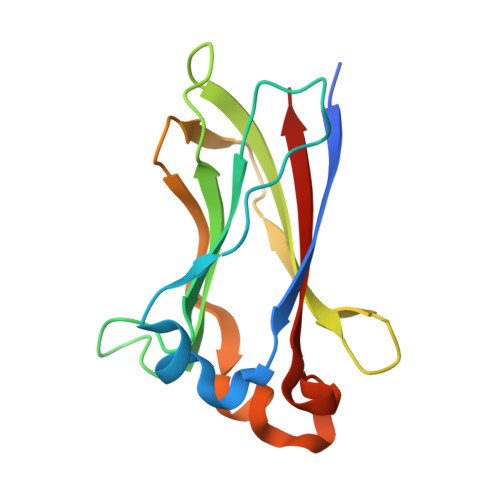
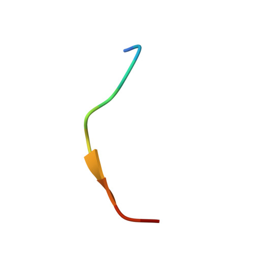
The Structure of the SPOP-Pdx1 Interface Reveals Insights into the Phosphorylation-Dependent Binding Regulation.

Ostertag, M.S.Messias, A.C.Sattler, M.Popowicz, G.M.

(2019) Structure 27: 327-334.e3

  • DOI: https://doi.org/10.1016/j.str.2018.10.005
  • Primary Citation of Related Structures:  
    6F8F, 6F8G

  • PubMed Abstract: 

    Pdx1 is a transcription factor crucial for development and maintenance of a functional pancreas. It regulates insulin expression and glucose homeostasis. SPOP is an E3-ubiquitin ligase adaptor protein that binds Pdx1, thus triggering its ubiquitination and proteasomal degradation. However, the underlying mechanisms are not well understood. Here, we present the crystal structure of the SPOP-Pdx1 complex. We show that Pdx1 residues 223-233 bind to SPOP MATH domain with low micromolar affinity. The interface is extended compared to other SPOP-client proteins. Previously, Pdx1 phosphorylation has been proposed to have a regulatory function. In this respect we show that phosphorylation lowers the affinity of Pdx1 to SPOP by isothermal titration calorimetry and nuclear magnetic resonance data. Our data provide insights into a critical protein-protein interaction that regulates cellular Pdx1 levels by SPOP-mediated decay. A reduction of Pdx1 levels in β cells is linked to apoptosis and considered a hallmark of type 2 diabetes.


  • Organizational Affiliation

    Institute of Structural Biology, Helmholtz Zentrum München, Ingolstädter Landstr. 1, Neuherberg 85764, Germany; Center for Integrated Protein Science Munich at Chair of Biomolecular NMR Spectroscopy, Department Chemie, Technische Universität München, Lichtenbergstrasse 4, Garching 85747, Germany.


Macromolecules
Find similar proteins by:  (by identity cutoff)  |  3D Structure
Entity ID: 1
MoleculeChains Sequence LengthOrganismDetailsImage
Speckle-type POZ proteinA [auth D]145Homo sapiensMutation(s): 0 
Gene Names: SPOP
UniProt & NIH Common Fund Data Resources
Find proteins for O43791 (Homo sapiens)
Explore O43791 
Go to UniProtKB:  O43791
PHAROS:  O43791
GTEx:  ENSG00000121067 
Entity Groups  
Sequence Clusters30% Identity50% Identity70% Identity90% Identity95% Identity100% Identity
UniProt GroupO43791
Sequence Annotations
Expand
  • Reference Sequence

Find similar proteins by:  Sequence   |   3D Structure  

Entity ID: 2
MoleculeChains Sequence LengthOrganismDetailsImage
Pancreas/duodenum homeobox protein 1B [auth G]11Homo sapiensMutation(s): 0 
UniProt & NIH Common Fund Data Resources
Find proteins for P52945 (Homo sapiens)
Explore P52945 
Go to UniProtKB:  P52945
PHAROS:  P52945
GTEx:  ENSG00000139515 
Entity Groups  
Sequence Clusters30% Identity50% Identity70% Identity90% Identity95% Identity100% Identity
UniProt GroupP52945
Sequence Annotations
Expand
  • Reference Sequence
Experimental Data & Validation

Experimental Data

  • Method: X-RAY DIFFRACTION
  • Resolution: 2.00 Å
  • R-Value Free: 0.274 
  • R-Value Work: 0.215 
  • R-Value Observed: 0.218 
  • Space Group: P 65 2 2
Unit Cell:
Length ( Å )Angle ( ˚ )
a = 53.925α = 90
b = 53.925β = 90
c = 207.41γ = 120
Software Package:
Software NamePurpose
REFMACrefinement
XDSdata reduction
XSCALEdata scaling
MOLREPphasing

Structure Validation

View Full Validation Report


Entry History 

Deposition Data

Revision History  (Full details and data files)

  • Version 1.0: 2018-11-28
    Type: Initial release
  • Version 1.1: 2019-02-13
    Changes: Data collection, Database references
  • Version 1.2: 2024-01-17
    Changes: Data collection, Database references, Refinement description