6E7K

Structure of the lipoprotein lipase GPIHBP1 complex that mediates plasma triglyceride hydrolysis


Experimental Data Snapshot

  • Method: X-RAY DIFFRACTION
  • Resolution: 2.80 Å
  • R-Value Free: 0.235 
  • R-Value Work: 0.195 
  • R-Value Observed: 0.197 

wwPDB Validation   3D Report Full Report


Ligand Structure Quality Assessment 


This is version 2.1 of the entry. See complete history


Literature

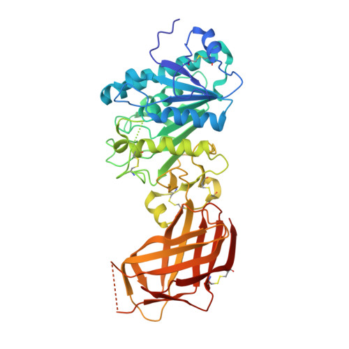
Structure of the lipoprotein lipase-GPIHBP1 complex that mediates plasma triglyceride hydrolysis.

Birrane, G.Beigneux, A.P.Dwyer, B.Strack-Logue, B.Kristensen, K.K.Francone, O.L.Fong, L.G.Mertens, H.D.T.Pan, C.Q.Ploug, M.Young, S.G.Meiyappan, M.

(2019) Proc Natl Acad Sci U S A 116: 1723-1732

  • DOI: https://doi.org/10.1073/pnas.1817984116
  • Primary Citation of Related Structures:  
    6E7K

  • PubMed Abstract: 

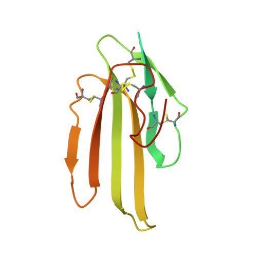
    Lipoprotein lipase (LPL) is responsible for the intravascular processing of triglyceride-rich lipoproteins. The LPL within capillaries is bound to GPIHBP1, an endothelial cell protein with a three-fingered LU domain and an N-terminal intrinsically disordered acidic domain. Loss-of-function mutations in LPL or GPIHBP1 cause severe hypertriglyceridemia (chylomicronemia), but structures for LPL and GPIHBP1 have remained elusive. Inspired by our recent discovery that GPIHBP1's acidic domain preserves LPL structure and activity, we crystallized an LPL-GPIHBP1 complex and solved its structure. GPIHBP1's LU domain binds to LPL's C-terminal domain, largely by hydrophobic interactions. Analysis of electrostatic surfaces revealed that LPL contains a large basic patch spanning its N- and C-terminal domains. GPIHBP1's acidic domain was not defined in the electron density map but was positioned to interact with LPL's large basic patch, providing a likely explanation for how GPIHBP1 stabilizes LPL. The LPL-GPIHBP1 structure provides insights into mutations causing chylomicronemia.


  • Organizational Affiliation

    Division of Experimental Medicine, Beth Israel Deaconess Medical Center, Boston, MA 02215.


Macromolecules
Find similar proteins by:  (by identity cutoff)  |  3D Structure
Entity ID: 1
MoleculeChains Sequence LengthOrganismDetailsImage
Lipoprotein lipase
A, B
448Homo sapiensMutation(s): 1 
Gene Names: LPLLIPD
EC: 3.1.1.34
UniProt & NIH Common Fund Data Resources
Find proteins for P06858 (Homo sapiens)
Explore P06858 
Go to UniProtKB:  P06858
PHAROS:  P06858
GTEx:  ENSG00000175445 
Entity Groups  
Sequence Clusters30% Identity50% Identity70% Identity90% Identity95% Identity100% Identity
UniProt GroupP06858
Sequence Annotations
Expand
  • Reference Sequence
Find similar proteins by:  (by identity cutoff)  |  3D Structure
Entity ID: 2
MoleculeChains Sequence LengthOrganismDetailsImage
Glycosylphosphatidylinositol-anchored high density lipoprotein-binding protein 1
C, D
131Homo sapiensMutation(s): 0 
Gene Names: GPIHBP1HBP1
UniProt & NIH Common Fund Data Resources
Find proteins for Q8IV16 (Homo sapiens)
Explore Q8IV16 
Go to UniProtKB:  Q8IV16
PHAROS:  Q8IV16
GTEx:  ENSG00000277494 
Entity Groups  
Sequence Clusters30% Identity50% Identity70% Identity90% Identity95% Identity100% Identity
UniProt GroupQ8IV16
Sequence Annotations
Expand
  • Reference Sequence
Oligosaccharides

Help

Entity ID: 3
MoleculeChains Length2D Diagram Glycosylation3D Interactions
beta-D-mannopyranose-(1-4)-2-acetamido-2-deoxy-beta-D-glucopyranose-(1-4)-2-acetamido-2-deoxy-beta-D-glucopyranose
E
3N-Glycosylation
Glycosylation Resources
GlyTouCan:  G15407YE
GlyCosmos:  G15407YE
GlyGen:  G15407YE
Entity ID: 4
MoleculeChains Length2D Diagram Glycosylation3D Interactions
beta-D-mannopyranose-(1-4)-2-acetamido-2-deoxy-beta-D-glucopyranose-(1-4)-[alpha-L-fucopyranose-(1-6)]2-acetamido-2-deoxy-beta-D-glucopyranose
F
4N-Glycosylation
Glycosylation Resources
GlyTouCan:  G32152BH
GlyCosmos:  G32152BH
GlyGen:  G32152BH
Entity ID: 5
MoleculeChains Length2D Diagram Glycosylation3D Interactions
2-acetamido-2-deoxy-beta-D-glucopyranose-(1-4)-[alpha-L-fucopyranose-(1-6)]2-acetamido-2-deoxy-beta-D-glucopyranose
G
3N-Glycosylation
Glycosylation Resources
GlyTouCan:  G21290RB
GlyCosmos:  G21290RB
GlyGen:  G21290RB
Experimental Data & Validation

Experimental Data

  • Method: X-RAY DIFFRACTION
  • Resolution: 2.80 Å
  • R-Value Free: 0.235 
  • R-Value Work: 0.195 
  • R-Value Observed: 0.197 
  • Space Group: P 21 21 2
Unit Cell:
Length ( Å )Angle ( ˚ )
a = 101.948α = 90
b = 153.206β = 90
c = 95.783γ = 90
Software Package:
Software NamePurpose
REFMACrefinement
HKL-2000data reduction
HKL-2000data scaling
PHASERphasing

Structure Validation

View Full Validation Report


Ligand Structure Quality Assessment 


Entry History 

Deposition Data

Revision History  (Full details and data files)

  • Version 1.0: 2018-12-19
    Type: Initial release
  • Version 1.1: 2019-01-02
    Changes: Data collection, Database references
  • Version 1.2: 2019-02-13
    Changes: Data collection, Database references
  • Version 2.0: 2020-07-29
    Type: Remediation
    Reason: Carbohydrate remediation
    Changes: Atomic model, Data collection, Derived calculations, Structure summary
  • Version 2.1: 2023-10-11
    Changes: Data collection, Database references, Derived calculations, Refinement description, Structure summary