6DF8

Kaiso (ZBTB33) zinc finger DNA binding domain in complex with the specific Kaiso binding sequence (KBS), pH 6.5


Experimental Data Snapshot

  • Method: X-RAY DIFFRACTION
  • Resolution: 2.54 Å
  • R-Value Free: 0.222 
  • R-Value Work: 0.179 
  • R-Value Observed: 0.184 

wwPDB Validation   3D Report Full Report


This is version 1.3 of the entry. See complete history


Literature

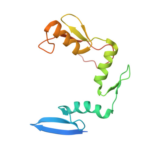
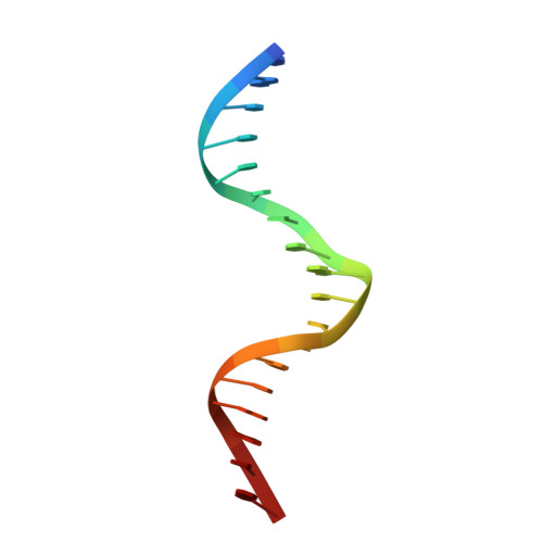
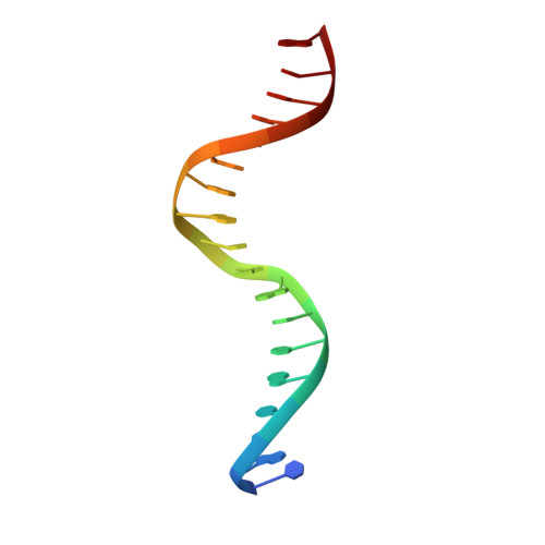
A conformational switch in the zinc finger protein Kaiso mediates differential readout of specific and methylated DNA sequences.

Nikolova, E.N.Stanfield, R.L.Dyson, H.J.Wright, P.E.

(2020) Biochemistry 

  • DOI: https://doi.org/10.1021/acs.biochem.0c00253
  • Primary Citation of Related Structures:  
    6DF5, 6DF8, 6DF9, 6DFA, 6DFB, 6DFC, 6V8U

  • PubMed Abstract: 

    Recognition of the epigenetic mark 5-methylcytosine (mC) at CpG sites in DNA has emerged as a novel function of many eukaryotic transcription factors (TFs). It remains unclear why the sequence specificity of these TFs differs for CpG-methylated motifs and consensus motifs. Here, we dissect the structural and dynamic basis for this differential DNA binding specificity in the human zinc finger TF Kaiso, which exhibits high affinity for two consecutive mCpG sites in variable contexts and also for a longer, sequence-specific Kaiso binding site (KBS). By integrating structural analysis and DNA binding studies with targeted protein mutagenesis and nucleotide substitutions, we identify distinct mechanisms for readout of methylated and KBS motifs by Kaiso. We show that a key glutamate residue (E535), critical for mCpG site recognition, adopts different conformations in complexes with specific and methylated DNA. These conformational differences, together with intrinsic variations in DNA flexibility and/or solvation at TpG versus mCpG sites, contribute to the different DNA affinity and sequence specificity. With methylated DNA, multiple direct contacts between E535 and the 5' mCpG site dominate the binding affinity, allowing for tolerance of different flanking DNA sequences. With KBS, Kaiso employs E535 as part of an indirect screen of the 5' flanking sequence, relying on key tyrosine-DNA interactions to stabilize an optimal DNA conformation and select against noncognate sites. These findings demonstrate how TFs use conformational adaptation and exploit variations in DNA flexibility to achieve distinct DNA readout outcomes and target a greater variety of regulatory and epigenetic sites than previously appreciated.


  • Organizational Affiliation

    Department of Integrative Structural and Computational Biology and Skaggs Institute of Chemical Biology, The Scripps Research Institute, 10550 North Torrey Pines Road, La Jolla, California 92037, United States.


Macromolecules

Find similar proteins by:  (by identity cutoff)  |  3D Structure
Entity ID: 1
MoleculeChains Sequence LengthOrganismDetailsImage
Transcriptional regulator Kaiso134Homo sapiensMutation(s): 0 
Gene Names: ZBTB33KAISOZNF348
UniProt & NIH Common Fund Data Resources
Find proteins for Q86T24 (Homo sapiens)
Explore Q86T24 
Go to UniProtKB:  Q86T24
PHAROS:  Q86T24
GTEx:  ENSG00000177485 
Entity Groups  
Sequence Clusters30% Identity50% Identity70% Identity90% Identity95% Identity100% Identity
UniProt GroupQ86T24
Sequence Annotations
Expand
  • Reference Sequence

Find similar nucleic acids by:  Sequence   |   3D Structure  

Entity ID: 2
MoleculeChains LengthOrganismImage
DNA (5'-D(*TP*GP*CP*TP*TP*CP*CP*TP*GP*CP*CP*AP*AP*TP*AP*AP*CP*G)-3')B [auth D]18Homo sapiens
Sequence Annotations
Expand
  • Reference Sequence

Find similar nucleic acids by:  Sequence   |   3D Structure  

Entity ID: 3
MoleculeChains LengthOrganismImage
DNA (5'-D(*CP*GP*TP*TP*AP*TP*TP*GP*GP*CP*AP*GP*GP*AP*AP*GP*CP*A)-3')C [auth E]18Homo sapiens
Sequence Annotations
Expand
  • Reference Sequence
Experimental Data & Validation

Experimental Data

  • Method: X-RAY DIFFRACTION
  • Resolution: 2.54 Å
  • R-Value Free: 0.222 
  • R-Value Work: 0.179 
  • R-Value Observed: 0.184 
  • Space Group: C 2 2 2
Unit Cell:
Length ( Å )Angle ( ˚ )
a = 44.371α = 90
b = 184.267β = 90
c = 104.938γ = 90
Software Package:
Software NamePurpose
PHENIXrefinement
HKL-2000data reduction
HKL-2000data scaling
PHENIXphasing

Structure Validation

View Full Validation Report


Entry History & Funding Information

Deposition Data


Funding OrganizationLocationGrant Number
National Institutes of Health/National Institute of General Medical Sciences (NIH/NIGMS)United StatesGM36643

Revision History  (Full details and data files)

  • Version 1.0: 2019-05-22
    Type: Initial release
  • Version 1.1: 2020-05-13
    Changes: Database references
  • Version 1.2: 2022-03-23
    Changes: Author supporting evidence, Database references
  • Version 1.3: 2023-10-11
    Changes: Data collection, Refinement description