6DCX

iASPP-PP-1c structure and targeting of p53


Experimental Data Snapshot

  • Method: X-RAY DIFFRACTION
  • Resolution: 3.41 Å
  • R-Value Free: 0.241 
  • R-Value Work: 0.187 
  • R-Value Observed: 0.190 

wwPDB Validation   3D Report Full Report


This is version 1.2 of the entry. See complete history


Literature

Flexible Tethering of ASPP Proteins Facilitates PP-1c Catalysis.

Zhou, Y.Millott, R.Kim, H.J.Peng, S.Edwards, R.A.Skene-Arnold, T.Hammel, M.Lees-Miller, S.P.Tainer, J.A.Holmes, C.F.B.Glover, J.N.M.

(2019) Structure 27: 1485-1496.e4

  • DOI: https://doi.org/10.1016/j.str.2019.07.012
  • Primary Citation of Related Structures:  
    6DCX

  • PubMed Abstract: 

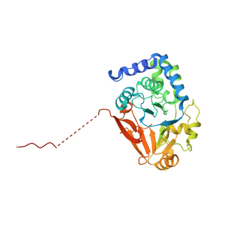
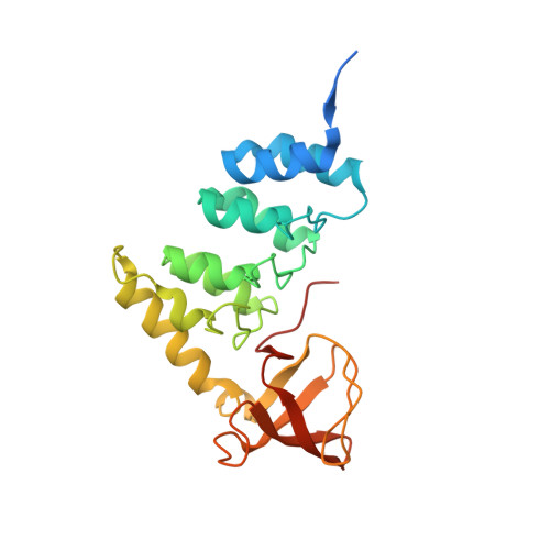
    ASPP (apoptosis-stimulating proteins of p53) proteins bind PP-1c (protein phosphatase 1) and regulate p53 impacting cancer cell growth and apoptosis. Here we determine the crystal structure of the oncogenic ASPP protein, iASPP, bound to PP-1c. The structure reveals a 1:1 complex that relies on interactions of the iASPP SILK and RVxF motifs with PP-1c, plus interactions of the PP-1c PxxPxR motif with the iASPP SH3 domain. Small-angle X-ray scattering analyses suggest that the crystal structure undergoes slow interconversion with more extended conformations in solution. We show that iASPP, and the tumor suppressor ASPP2, enhance the catalytic activity of PP-1c against the small-molecule substrate, pNPP as well as p53. The combined results suggest that PxxPxR binding to iASPP SH3 domain is critical for complex formation, and that the modular ASPP-PP-1c interface provides dynamic flexibility that enables functional binding and dephosphorylation of p53 and other diverse protein substrates.


  • Organizational Affiliation

    Department of Biochemistry, University of Alberta, Edmonton, AB T6G 2H7, Canada.


Macromolecules
Find similar proteins by:  (by identity cutoff)  |  3D Structure
Entity ID: 1
MoleculeChains Sequence LengthOrganismDetailsImage
Serine/threonine-protein phosphatase PP1-alpha catalytic subunit
A, B
339Homo sapiensMutation(s): 0 
Gene Names: PPP1CAPPP1A
EC: 3.1.3.16
UniProt & NIH Common Fund Data Resources
Find proteins for P62136 (Homo sapiens)
Explore P62136 
Go to UniProtKB:  P62136
PHAROS:  P62136
GTEx:  ENSG00000172531 
Entity Groups  
Sequence Clusters30% Identity50% Identity70% Identity90% Identity95% Identity100% Identity
UniProt GroupP62136
Sequence Annotations
Expand
  • Reference Sequence
Find similar proteins by:  (by identity cutoff)  |  3D Structure
Entity ID: 2
MoleculeChains Sequence LengthOrganismDetailsImage
RelA-associated inhibitor
C, D
228Homo sapiensMutation(s): 0 
Gene Names: PPP1R13LIASPPNKIP1PPP1R13BLRAI
UniProt & NIH Common Fund Data Resources
Find proteins for Q8WUF5 (Homo sapiens)
Explore Q8WUF5 
Go to UniProtKB:  Q8WUF5
PHAROS:  Q8WUF5
GTEx:  ENSG00000104881 
Entity Groups  
Sequence Clusters30% Identity50% Identity70% Identity90% Identity95% Identity100% Identity
UniProt GroupQ8WUF5
Sequence Annotations
Expand
  • Reference Sequence
Experimental Data & Validation

Experimental Data

  • Method: X-RAY DIFFRACTION
  • Resolution: 3.41 Å
  • R-Value Free: 0.241 
  • R-Value Work: 0.187 
  • R-Value Observed: 0.190 
  • Space Group: P 41 21 2
Unit Cell:
Length ( Å )Angle ( ˚ )
a = 135.462α = 90
b = 135.462β = 90
c = 165.39γ = 90
Software Package:
Software NamePurpose
HKL-2000data collection
HKL-2000data scaling
PHASERphasing
PHENIXrefinement
PDB_EXTRACTdata extraction
HKL-2000data reduction

Structure Validation

View Full Validation Report


Entry History 

Deposition Data

Revision History  (Full details and data files)

  • Version 1.0: 2019-05-15
    Type: Initial release
  • Version 1.1: 2019-11-27
    Changes: Database references
  • Version 1.2: 2023-10-11
    Changes: Data collection, Database references, Refinement description