6C0T

Crystal structure of cGMP-dependent protein kinase Ialpha (PKG Ialpha) catalytic domain bound with N46


Experimental Data Snapshot

  • Method: X-RAY DIFFRACTION
  • Resolution: 1.98 Å
  • R-Value Free: 0.244 
  • R-Value Work: 0.207 
  • R-Value Observed: 0.210 

wwPDB Validation   3D Report Full Report


Ligand Structure Quality Assessment 


This is version 1.3 of the entry. See complete history


Literature

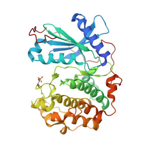
Structural basis for selective inhibition of human PKG I alpha by the balanol-like compound N46.

Qin, L.Sankaran, B.Aminzai, S.Casteel, D.E.Kim, C.

(2018) J Biol Chem 293: 10985-10992

  • DOI: https://doi.org/10.1074/jbc.RA118.002427
  • Primary Citation of Related Structures:  
    6C0T, 6C0U

  • PubMed Abstract: 

    Activation of protein kinase G (PKG) Iα in nociceptive neurons induces long-term hyperexcitability that causes chronic pain. Recently, a derivative of the fungal metabolite balanol, N46, has been reported to inhibit PKG Iα with high potency and selectivity and attenuate thermal hyperalgesia and osteoarthritic pain. Here we determined co-crystal structures of the PKG Iα C-domain and cAMP-dependent protein kinase (PKA) Cα, each bound with N46, at 1.98 Å and 2.65 Å, respectively. N46 binds the active site with its external phenyl ring, specifically interacting with the glycine-rich loop and the αC helix. Phe-371 at the PKG Iα glycine-rich loop is oriented parallel to the phenyl ring of N46, forming a strong π-stacking interaction, whereas the analogous Phe-54 in PKA Cα rotates 30° and forms a weaker interaction. Structural comparison revealed that steric hindrance between the preceding Ser-53 and the propoxy group of the phenyl ring may explain the weaker interaction with PKA Cα. The analogous Gly-370 in PKG Iα, however, causes little steric hindrance with Phe-371. Moreover, Ile-406 on the αC helix forms a hydrophobic interaction with N46 whereas its counterpart in PKA, Thr-88, does not. Substituting these residues in PKG Iα with those in PKA Cα increases the IC 50 values for N46, whereas replacing these residues in PKA Cα with those in PKG Iα reduces the IC 50 , consistent with our structural findings. In conclusion, our results explain the structural basis for N46-mediated selective inhibition of human PKG Iα and provide a starting point for structure-guided design of selective PKG Iα inhibitors.


  • Organizational Affiliation

    From the Verna and Marrs McLean Department of Biochemistry and Molecular Biology, Baylor College of Medicine, Houston, Texas 77030.


Macromolecules
Find similar proteins by:  (by identity cutoff)  |  3D Structure
Entity ID: 1
MoleculeChains Sequence LengthOrganismDetailsImage
cGMP-dependent protein kinase 1347Homo sapiensMutation(s): 0 
Gene Names: PRKG1PRKG1BPRKGR1APRKGR1B
EC: 2.7.11.12
UniProt & NIH Common Fund Data Resources
Find proteins for Q13976 (Homo sapiens)
Explore Q13976 
Go to UniProtKB:  Q13976
PHAROS:  Q13976
GTEx:  ENSG00000185532 
Entity Groups  
Sequence Clusters30% Identity50% Identity70% Identity90% Identity95% Identity100% Identity
UniProt GroupQ13976
Sequence Annotations
Expand
  • Reference Sequence
Small Molecules
Ligands 3 Unique
IDChains Name / Formula / InChI Key2D Diagram3D Interactions
EE4
Query on EE4

Download Ideal Coordinates CCD File 
B [auth A]N-[(3R,4R)-4-{[4-(2-fluoro-3-methoxy-6-propoxybenzene-1-carbonyl)benzene-1-carbonyl]amino}pyrrolidin-3-yl]-1H-indazole-5-carboxamide
C30 H30 F N5 O5
AYXNTJKOINXKAJ-DHIUTWEWSA-N
PGE
Query on PGE

Download Ideal Coordinates CCD File 
D [auth A]TRIETHYLENE GLYCOL
C6 H14 O4
ZIBGPFATKBEMQZ-UHFFFAOYSA-N
DMS
Query on DMS

Download Ideal Coordinates CCD File 
C [auth A]DIMETHYL SULFOXIDE
C2 H6 O S
IAZDPXIOMUYVGZ-UHFFFAOYSA-N
Modified Residues  1 Unique
IDChains TypeFormula2D DiagramParent
TPO
Query on TPO
A
L-PEPTIDE LINKINGC4 H10 N O6 PTHR
Binding Affinity Annotations 
IDSourceBinding Affinity
EE4 Binding MOAD:  6C0T IC50: 43.1 (nM) from 1 assay(s)
Experimental Data & Validation

Experimental Data

  • Method: X-RAY DIFFRACTION
  • Resolution: 1.98 Å
  • R-Value Free: 0.244 
  • R-Value Work: 0.207 
  • R-Value Observed: 0.210 
  • Space Group: P 42
Unit Cell:
Length ( Å )Angle ( ˚ )
a = 85.73α = 90
b = 85.73β = 90
c = 50.664γ = 90
Software Package:
Software NamePurpose
PHENIXrefinement
iMOSFLMdata reduction
Aimlessdata scaling
PHENIXphasing

Structure Validation

View Full Validation Report


Ligand Structure Quality Assessment 


Entry History & Funding Information

Deposition Data


Funding OrganizationLocationGrant Number
National Institutes of Health/National Institute of General Medical Sciences (NIH/NIGMS)United States5R01GM090161-08

Revision History  (Full details and data files)

  • Version 1.0: 2018-05-30
    Type: Initial release
  • Version 1.1: 2018-07-25
    Changes: Data collection, Database references
  • Version 1.2: 2020-01-01
    Changes: Author supporting evidence
  • Version 1.3: 2023-10-04
    Changes: Data collection, Database references, Refinement description