6BWY

DNA substrate selection by APOBEC3G


Experimental Data Snapshot

  • Method: X-RAY DIFFRACTION
  • Resolution: 2.90 Å
  • R-Value Free: 0.287 
  • R-Value Work: 0.234 
  • R-Value Observed: 0.237 

wwPDB Validation   3D Report Full Report


This is version 1.1 of the entry. See complete history


Literature

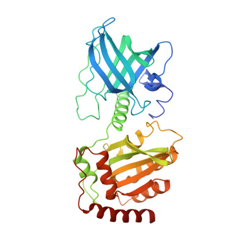
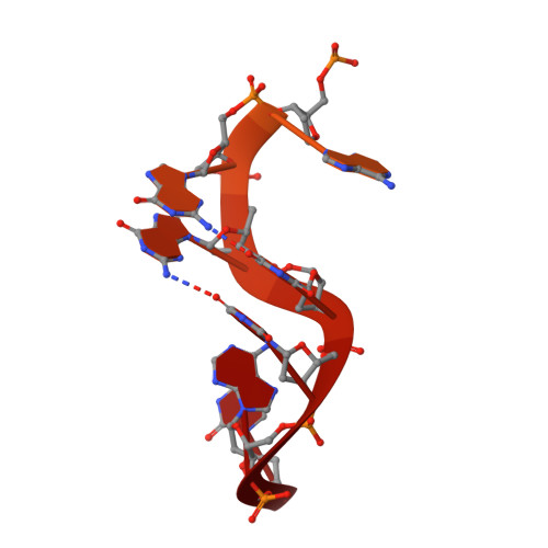
Insights into DNA substrate selection by APOBEC3G from structural, biochemical, and functional studies.

Ziegler, S.J.Liu, C.Landau, M.Buzovetsky, O.Desimmie, B.A.Zhao, Q.Sasaki, T.Burdick, R.C.Pathak, V.K.Anderson, K.S.Xiong, Y.

(2018) PLoS One 13: e0195048-e0195048

  • DOI: https://doi.org/10.1371/journal.pone.0195048
  • Primary Citation of Related Structures:  
    6BWY

  • PubMed Abstract: 

    Human apolipoprotein B mRNA-editing enzyme-catalytic polypeptide-like 3 (A3) proteins are a family of cytidine deaminases that catalyze the conversion of deoxycytidine (dC) to deoxyuridine (dU) in single-stranded DNA (ssDNA). A3 proteins act in the innate immune response to viral infection by mutating the viral ssDNA. One of the most well-studied human A3 family members is A3G, which is a potent inhibitor of HIV-1. Each A3 protein prefers a specific substrate sequence for catalysis-for example, A3G deaminates the third dC in the CCCA sequence motif. However, the interaction between A3G and ssDNA is difficult to characterize due to poor solution behavior of the full-length protein and loss of DNA affinity of the truncated protein. Here, we present a novel DNA-anchoring fusion strategy using the protection of telomeres protein 1 (Pot1) which has nanomolar affinity for ssDNA, with which we captured an A3G-ssDNA interaction. We crystallized a non-preferred adenine in the -1 nucleotide-binding pocket of A3G. The structure reveals a unique conformation of the catalytic site loops that sheds light onto how the enzyme scans substrate in the -1 pocket. Furthermore, our biochemistry and virology studies provide evidence that the nucleotide-binding pockets on A3G influence each other in selecting the preferred DNA substrate. Together, the results provide insights into the mechanism by which A3G selects and deaminates its preferred substrates and help define how A3 proteins are tailored to recognize specific DNA sequences. This knowledge contributes to a better understanding of the mechanism of DNA substrate selection by A3G, as well as A3G antiviral activity against HIV-1.


  • Organizational Affiliation

    Department of Molecular Biophysics and Biochemistry, Yale University, New Haven, Connecticut, United States of America.


Macromolecules

Find similar proteins by:  (by identity cutoff)  |  3D Structure
Entity ID: 2
MoleculeChains Sequence LengthOrganismDetailsImage
Protection of telomeres protein 1, DNA dC->dU-editing enzyme APOBEC-3G fusionE [auth A],
F [auth B],
G [auth E],
H [auth G]
361Schizosaccharomyces pombe 972h-Homo sapiens
This entity is chimeric
Mutation(s): 5 
Gene Names: pot1SPAC26H5.06APOBEC3GMDS019
EC: 3.5.4
UniProt & NIH Common Fund Data Resources
Find proteins for O13988 (Schizosaccharomyces pombe (strain 972 / ATCC 24843))
Explore O13988 
Go to UniProtKB:  O13988
Find proteins for Q9HC16 (Homo sapiens)
Explore Q9HC16 
Go to UniProtKB:  Q9HC16
PHAROS:  Q9HC16
GTEx:  ENSG00000239713 
Entity Groups  
Sequence Clusters30% Identity50% Identity70% Identity90% Identity95% Identity100% Identity
UniProt GroupsO13988Q9HC16
Sequence Annotations
Expand
  • Reference Sequence
Find similar nucleic acids by:  (by identity cutoff)  |  3D Structure
Entity ID: 1
MoleculeChains LengthOrganismImage
DNA (30-MER)A [auth I],
B [auth C],
C [auth D],
D [auth F]
30synthetic construct
Sequence Annotations
Expand
  • Reference Sequence
Small Molecules
Ligands 2 Unique
IDChains Name / Formula / InChI Key2D Diagram3D Interactions
PO4
Query on PO4

Download Ideal Coordinates CCD File 
AA [auth G]
I
J [auth C]
K [auth D]
L [auth F]
AA [auth G],
I,
J [auth C],
K [auth D],
L [auth F],
N [auth A],
O [auth A],
P [auth A],
R [auth B],
S [auth B],
U [auth E],
V [auth E],
W [auth E],
Y [auth G],
Z [auth G]
PHOSPHATE ION
O4 P
NBIIXXVUZAFLBC-UHFFFAOYSA-K
ZN
Query on ZN

Download Ideal Coordinates CCD File 
M [auth A],
Q [auth B],
T [auth E],
X [auth G]
ZINC ION
Zn
PTFCDOFLOPIGGS-UHFFFAOYSA-N
Experimental Data & Validation

Experimental Data

  • Method: X-RAY DIFFRACTION
  • Resolution: 2.90 Å
  • R-Value Free: 0.287 
  • R-Value Work: 0.234 
  • R-Value Observed: 0.237 
  • Space Group: P 43
Unit Cell:
Length ( Å )Angle ( ˚ )
a = 79.072α = 90
b = 79.072β = 90
c = 266.411γ = 90
Software Package:
Software NamePurpose
REFMACrefinement
HKL-2000data reduction
HKL-2000data scaling
PHASERphasing

Structure Validation

View Full Validation Report


Entry History 

Deposition Data

Revision History  (Full details and data files)

  • Version 1.0: 2018-04-18
    Type: Initial release
  • Version 1.1: 2023-10-04
    Changes: Data collection, Database references, Refinement description