Carboxylic Acid Derivatives of Amlexanox Display Enhanced Potency toward TBK1 and IKKepsilonand Reveal Mechanisms for Selective Inhibition.
Beyett, T.S., Gan, X., Reilly, S.M., Chang, L., Gomez, A.V., Saltiel, A.R., Showalter, H.D., Tesmer, J.J.G.(2018) Mol Pharmacol 94: 1210-1219
- PubMed: 30082428
- DOI: https://doi.org/10.1124/mol.118.112185
- Primary Citation of Related Structures:
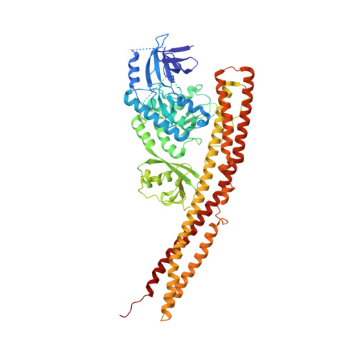
5W5V, 6BNY, 6BOD, 6BOE - PubMed Abstract:
Chronic low-grade inflammation is a hallmark of obesity, which is a risk factor for the development of type 2 diabetes. The drug amlexanox inhibits I κ B kinase ε (IKK ε ) and TANK binding kinase 1 (TBK1) to promote energy expenditure and improve insulin sensitivity. Clinical studies have demonstrated efficacy in a subset of diabetic patients with underlying adipose tissue inflammation, albeit with moderate potency, necessitating the need for improved analogs. Herein we report crystal structures of TBK1 in complex with amlexanox and a series of analogs that modify its carboxylic acid moiety. Removal of the carboxylic acid or mutation of the adjacent Thr156 residue significantly reduces potency toward TBK1, whereas conversion to a short amide or ester nearly abolishes the inhibitory effects. IKK ε is less affected by these modifications, possibly due to variation in its hinge that allows for increased conformational plasticity. Installation of a tetrazole carboxylic acid bioisostere improved potency to 200 and 400 nM toward IKK ε and TBK1, respectively. Despite improvements in the in vitro potency, no analog produced a greater response in adipocytes than amlexanox, perhaps because of altered absorption and distribution. The structure-activity relationships and cocrystal structures described herein will aid in future structure-guided inhibitor development using the amlexanox pharmacophore for the treatment of obesity and type 2 diabetes.
Organizational Affiliation:
Program in Chemical Biology (T.S.B.), Life Sciences Institute (T.S.B., L.C., J.J.G.T.), Departments of Medicinal Chemistry (X.G., H.D.S., J.J.G.T.), Pharmacology (J.J.G.T.), Biological Chemistry (J.J.G.T.), and Vahlteich Medicinal Chemistry Core, College of Pharmacy (X.G., H.D.S.), University of Michigan, Ann Arbor, Michigan; Institute for Diabetes and Metabolic Health (S.M.R., A.V.G., A.R.S.), Departments of Medicine (S.M.R., A.R.S.) and Pharmacology (A.V.G., A.R.S.), University of California, San Diego, La Jolla, California; and Departments of Biological Sciences and of Medicinal Chemistry and Molecular Pharmacology, Purdue University, West Lafayette, Indiana (J.J.G.T.).