6A9Y

The crystal structure of Mu homology domain of SGIP1


Experimental Data Snapshot

  • Method: X-RAY DIFFRACTION
  • Resolution: 2.70 Å
  • R-Value Free: 0.266 
  • R-Value Work: 0.198 
  • R-Value Observed: 0.202 

wwPDB Validation   3D Report Full Report


This is version 1.3 of the entry. See complete history


Literature

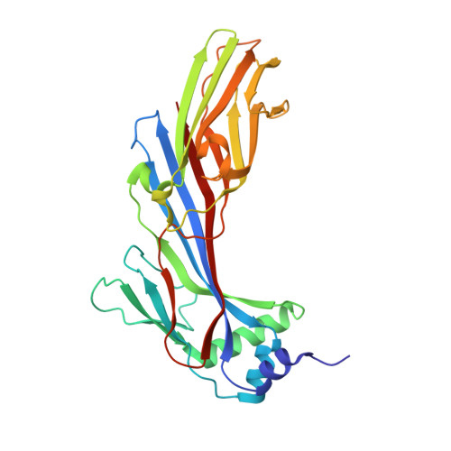
SGIP1 dimerizes via intermolecular disulfide bond in mu HD domain during cellular endocytosis.

Zhang, Y.Feng, Y.Xin, Y.Liu, X.

(2018) Biochem Biophys Res Commun 505: 99-105

  • DOI: https://doi.org/10.1016/j.bbrc.2018.09.075
  • Primary Citation of Related Structures:  
    6A9Y

  • PubMed Abstract: 

    Along with its homologs FCHo1 and FCHo2, SGIP1 plays an important role in clathrin-mediated endocytosis. The highly conserved C-terminal μHD domains in these proteins are the critical regions interacting with adapter molecules such as Eps15. The crystal structure of μHD domain of SGIP1 has been reported previously. In this study, we found that μHD domain of SGIP1 is capable of forming a stable dimer by an intermolecular disulfide bond formed by C632 in our crystal structure. The mutational study of C632 revealed that this residue is important for the function of SGIP1 during cellular endocytosis. Our study revealed a new dimerization and/or oligomerization manner in theses adaptor proteins, which is a critical prerequisite for their proper function.


  • Organizational Affiliation

    State Key Laboratory of Medicinal Chemical Biology, Department of Biochemistry and Molecular Biology, College of Life Sciences, Nankai University, Tianjin, 300071, China.


Macromolecules
Find similar proteins by:  (by identity cutoff)  |  3D Structure
Entity ID: 1
MoleculeChains Sequence LengthOrganismDetailsImage
SH3-containing GRB2-like protein 3-interacting protein 1284Homo sapiensMutation(s): 0 
Gene Names: SGIP1
UniProt & NIH Common Fund Data Resources
Find proteins for Q9BQI5 (Homo sapiens)
Explore Q9BQI5 
Go to UniProtKB:  Q9BQI5
PHAROS:  Q9BQI5
GTEx:  ENSG00000118473 
Entity Groups  
Sequence Clusters30% Identity50% Identity70% Identity90% Identity95% Identity100% Identity
UniProt GroupQ9BQI5
Sequence Annotations
Expand
  • Reference Sequence
Experimental Data & Validation

Experimental Data

  • Method: X-RAY DIFFRACTION
  • Resolution: 2.70 Å
  • R-Value Free: 0.266 
  • R-Value Work: 0.198 
  • R-Value Observed: 0.202 
  • Space Group: P 21 21 2
Unit Cell:
Length ( Å )Angle ( ˚ )
a = 70.073α = 90
b = 132.308β = 90
c = 40.672γ = 90
Software Package:
Software NamePurpose
PHENIXrefinement
HKL-2000data reduction
HKL-2000data scaling
PHENIXphasing

Structure Validation

View Full Validation Report


Entry History 

Deposition Data

Revision History  (Full details and data files)

  • Version 1.0: 2018-09-26
    Type: Initial release
  • Version 1.1: 2018-10-10
    Changes: Data collection, Database references, Structure summary
  • Version 1.2: 2018-10-24
    Changes: Data collection, Database references, Structure summary
  • Version 1.3: 2023-11-22
    Changes: Data collection, Database references, Refinement description