5YBU

Structure of the KANK1 ankyrin domain in complex with KIF21A peptide


Experimental Data Snapshot

  • Method: X-RAY DIFFRACTION
  • Resolution: 1.89 Å
  • R-Value Free: 0.253 
  • R-Value Work: 0.222 
  • R-Value Observed: 0.225 

wwPDB Validation   3D Report Full Report


This is version 1.3 of the entry. See complete history


Literature

Structural basis for the recognition of kinesin family member 21A (KIF21A) by the ankyrin domains of KANK1 and KANK2 proteins.

Guo, Q.Liao, S.Zhu, Z.Li, Y.Li, F.Xu, C.

(2018) J Biol Chem 293: 557-566

  • DOI: https://doi.org/10.1074/jbc.M117.817494
  • Primary Citation of Related Structures:  
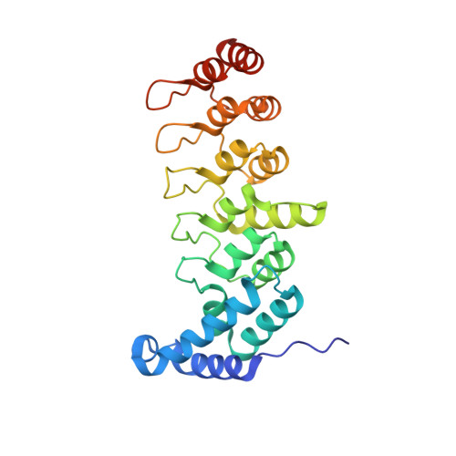
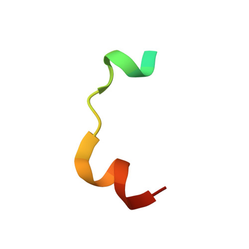
    5YBJ, 5YBU, 5YBV

  • PubMed Abstract: 

    A well-controlled microtubule organization is essential for intracellular transport, cytoskeleton maintenance, and cell development. KN motif and ankyrin repeat domain-containing protein 1 (KANK1), a member of KANK family, recruits kinesin family member 21A (KIF21A) to the cell cortex to control microtubule growth via its C-terminal ankyrin domain. However, how the KANK1 ankyrin domain recognizes KIF21A and whether other KANK proteins can also bind KIF21A remain unknown. Here, using a combination of structural, site-directed mutagenesis, and biochemical studies, we found that a stretch of ∼22 amino acids in KIF21A is sufficient for binding to KANK1 and its close homolog KANK2. We further solved the complex structure of the KIF21A peptide with either the KANK1 ankyrin domain or the KANK2 ankyrin domain. In each complex, KIF21A is recognized by two distinct pockets of the ankyrin domain and adopts helical conformations upon binding to the ankyrin domain. The elucidated KANK structures may advance our understanding of the role of KANK1 as a scaffolding molecule in controlling microtubule growth at the cell periphery.


  • Organizational Affiliation

    From the Hefei National Laboratory for Physical Sciences at the Microscale and School of Life Sciences, University of Science and Technology of China, Hefei 230027, China and.


Macromolecules
Find similar proteins by:  (by identity cutoff)  |  3D Structure
Entity ID: 1
MoleculeChains Sequence LengthOrganismDetailsImage
KN motif and ankyrin repeat domain-containing protein 1253Homo sapiensMutation(s): 0 
Gene Names: KANK1ANKRD15KANKKIAA0172
UniProt & NIH Common Fund Data Resources
Find proteins for Q14678 (Homo sapiens)
Explore Q14678 
Go to UniProtKB:  Q14678
PHAROS:  Q14678
GTEx:  ENSG00000107104 
Entity Groups  
Sequence Clusters30% Identity50% Identity70% Identity90% Identity95% Identity100% Identity
UniProt GroupQ14678
Sequence Annotations
Expand
  • Reference Sequence

Find similar proteins by:  Sequence   |   3D Structure  

Entity ID: 2
MoleculeChains Sequence LengthOrganismDetailsImage
Kinesin-like protein KIF21A22Homo sapiensMutation(s): 0 
Gene Names: KIF21AKIAA1708KIF2
UniProt & NIH Common Fund Data Resources
Find proteins for Q7Z4S6 (Homo sapiens)
Explore Q7Z4S6 
Go to UniProtKB:  Q7Z4S6
PHAROS:  Q7Z4S6
GTEx:  ENSG00000139116 
Entity Groups  
Sequence Clusters30% Identity50% Identity70% Identity90% Identity95% Identity100% Identity
UniProt GroupQ7Z4S6
Sequence Annotations
Expand
  • Reference Sequence
Experimental Data & Validation

Experimental Data

  • Method: X-RAY DIFFRACTION
  • Resolution: 1.89 Å
  • R-Value Free: 0.253 
  • R-Value Work: 0.222 
  • R-Value Observed: 0.225 
  • Space Group: P 21 21 21
Unit Cell:
Length ( Å )Angle ( ˚ )
a = 37.648α = 90
b = 51.624β = 90
c = 137.609γ = 90
Software Package:
Software NamePurpose
PHENIXrefinement
HKL-2000data processing
HKL-2000data scaling
PHENIXphasing

Structure Validation

View Full Validation Report


Entry History 

Deposition Data

Revision History  (Full details and data files)

  • Version 1.0: 2017-12-06
    Type: Initial release
  • Version 1.1: 2017-12-13
    Changes: Database references
  • Version 1.2: 2018-01-24
    Changes: Database references
  • Version 1.3: 2023-11-22
    Changes: Data collection, Database references, Refinement description