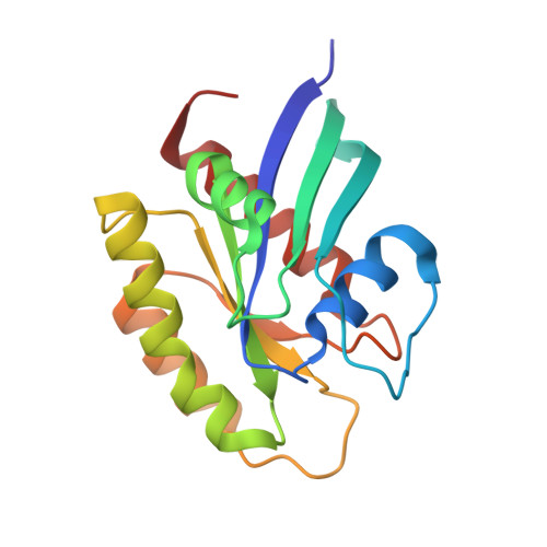
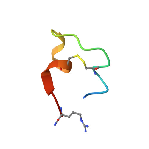
5XCO

Crystal structure of human K-Ras G12D Mutant in complex with GDP and Cyclic Inhibitory Peptide


Experimental Data Snapshot

  • Method: X-RAY DIFFRACTION
  • Resolution: 1.25 Å
  • R-Value Free: 0.194 
  • R-Value Work: 0.171 
  • R-Value Observed: 0.172 

wwPDB Validation   3D Report Full Report


Ligand Structure Quality Assessment 


This is version 1.2 of the entry. See complete history


Literature

Crystal Structure of a Human K-Ras G12D Mutant in Complex with GDP and the Cyclic Inhibitory Peptide KRpep-2d

Sogabe, S.Kamada, Y.Miwa, M.Niida, A.Sameshima, T.Kamaura, M.Yonemori, K.Sasaki, S.Sakamoto, J.Sakamoto, K.

(2017) ACS Med Chem Lett 8: 732-736

  • DOI: https://doi.org/10.1021/acsmedchemlett.7b00128
  • Primary Citation of Related Structures:  
    5XCO

  • PubMed Abstract: 

    The Ras proteins play roles in cell differentiation, proliferation, and survival. Aberrant signaling through Ras-mediated pathways in tumor cells occurs as a result of several types of mutational damage, which most frequently affects the amino acids G12, G13, and Q61. Recently, KRpep-2d was identified as a K-Ras(G12D) selective inhibitory peptide against the G12D mutant of K-Ras, which is a key member of the Ras protein family and an attractive cancer therapeutic target. In this study, the crystal structure of the human K-Ras(G12D) mutant was determined in complex with GDP and KRpep-2d at 1.25 Å resolution. This structure revealed that the peptide binds near Switch II and allosterically blocks protein-protein interactions with the guanine nucleotide exchange factor. This discovery of a unique binding pocket provides valuable information that will facilitate the design of direct Ras inhibitors.


  • Organizational Affiliation

    Pharmaceutical Research Division, Takeda Pharmaceutical Company Limited, 26-1, Muraoka-Higashi 2-chome, Fujisawa, Kanagawa 251-8555, Japan.


Macromolecules
Find similar proteins by:  (by identity cutoff)  |  3D Structure
Entity ID: 1
MoleculeChains Sequence LengthOrganismDetailsImage
GTPase KRas171Homo sapiensMutation(s): 1 
Gene Names: KRASKRAS2RASK2
UniProt & NIH Common Fund Data Resources
Find proteins for P01116 (Homo sapiens)
Explore P01116 
Go to UniProtKB:  P01116
PHAROS:  P01116
GTEx:  ENSG00000133703 
Entity Groups  
Sequence Clusters30% Identity50% Identity70% Identity90% Identity95% Identity100% Identity
UniProt GroupP01116
Sequence Annotations
Expand
  • Reference Sequence

Find similar proteins by:  Sequence   |   3D Structure  

Entity ID: 2
MoleculeChains Sequence LengthOrganismDetailsImage
ACE-ARG-ARG-ARG-ARG-CYS-PRO-LEU-TYR-ILE-SER-TYR-ASP-PRO-VAL-CYS-ARG-ARG-ARG-ARG-NH221Escherichia phage T7Mutation(s): 0 
Entity Groups  
Sequence Clusters30% Identity50% Identity70% Identity90% Identity95% Identity100% Identity
Sequence Annotations
Expand
  • Reference Sequence
Small Molecules
Ligands 2 Unique
IDChains Name / Formula / InChI Key2D Diagram3D Interactions
GDP
Query on GDP

Download Ideal Coordinates CCD File 
C [auth A]GUANOSINE-5'-DIPHOSPHATE
C10 H15 N5 O11 P2
QGWNDRXFNXRZMB-UUOKFMHZSA-N
EDO
Query on EDO

Download Ideal Coordinates CCD File 
D [auth A]1,2-ETHANEDIOL
C2 H6 O2
LYCAIKOWRPUZTN-UHFFFAOYSA-N
Experimental Data & Validation

Experimental Data

  • Method: X-RAY DIFFRACTION
  • Resolution: 1.25 Å
  • R-Value Free: 0.194 
  • R-Value Work: 0.171 
  • R-Value Observed: 0.172 
  • Space Group: P 31 2 1
Unit Cell:
Length ( Å )Angle ( ˚ )
a = 51.529α = 90
b = 51.529β = 90
c = 129.741γ = 120
Software Package:
Software NamePurpose
REFMACrefinement
HKL-2000data reduction
SCALEPACKdata scaling
PHASERphasing

Structure Validation

View Full Validation Report


Ligand Structure Quality Assessment 


Entry History 

Deposition Data

Revision History  (Full details and data files)

  • Version 1.0: 2017-05-24
    Type: Initial release
  • Version 1.1: 2017-08-09
    Changes: Database references
  • Version 1.2: 2023-11-22
    Changes: Data collection, Database references, Refinement description