5W6E

PDE1b complexed with compound 3S


Experimental Data Snapshot

  • Method: X-RAY DIFFRACTION
  • Resolution: 1.90 Å
  • R-Value Free: 0.206 
  • R-Value Work: 0.180 
  • R-Value Observed: 0.182 

wwPDB Validation   3D Report Full Report


Ligand Structure Quality Assessment 


This is version 1.2 of the entry. See complete history


Literature

Discovery of Potent and Selective Periphery-Restricted Quinazoline Inhibitors of the Cyclic Nucleotide Phosphodiesterase PDE1.

Humphrey, J.M.Movsesian, M.Am Ende, C.W.Becker, S.L.Chappie, T.A.Jenkinson, S.Liras, J.L.Liras, S.Orozco, C.Pandit, J.Vajdos, F.F.Vandeput, F.Yang, E.Menniti, F.S.

(2018) J Med Chem 61: 4635-4640

  • DOI: https://doi.org/10.1021/acs.jmedchem.8b00374
  • Primary Citation of Related Structures:  
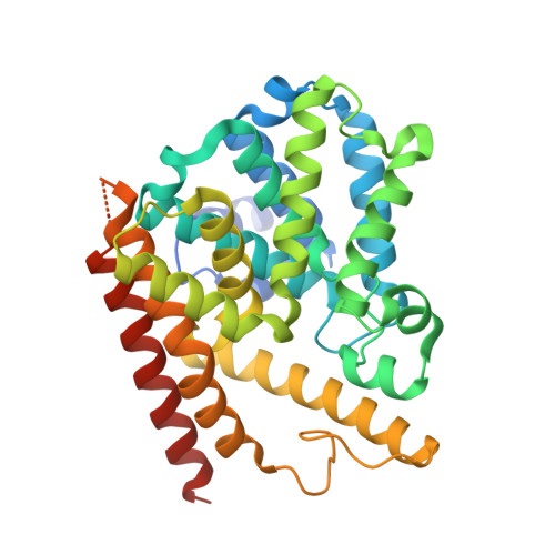
    5W6E

  • PubMed Abstract: 

    We disclose the discovery and X-ray cocrystal data of potent, selective quinazoline inhibitors of PDE1. Inhibitor ( S)-3 readily attains free plasma concentrations above PDE1 IC 50 values and has restricted brain access. The racemic compound 3 inhibits >75% of PDE hydrolytic activity in soluble samples of human myocardium, consistent with heightened PDE1 activity in this tissue. These compounds represent promising new tools to probe the value of PDE1 inhibition in the treatment of cardiovascular disease.


  • Organizational Affiliation

    Pfizer World Wide Research and Development , Eastern Point Road , Groton , Connecticut 06340 , United States.


Macromolecules
Find similar proteins by:  (by identity cutoff)  |  3D Structure
Entity ID: 1
MoleculeChains Sequence LengthOrganismDetailsImage
Calcium/calmodulin-dependent 3',5'-cyclic nucleotide phosphodiesterase 1B369Homo sapiensMutation(s): 0 
Gene Names: PDE1BPDE1B1PDES1B
EC: 3.1.4.17
UniProt & NIH Common Fund Data Resources
Find proteins for Q01064 (Homo sapiens)
Explore Q01064 
Go to UniProtKB:  Q01064
PHAROS:  Q01064
GTEx:  ENSG00000123360 
Entity Groups  
Sequence Clusters30% Identity50% Identity70% Identity90% Identity95% Identity100% Identity
UniProt GroupQ01064
Sequence Annotations
Expand
  • Reference Sequence
Small Molecules
Ligands 3 Unique
IDChains Name / Formula / InChI Key2D Diagram3D Interactions
0NY
Query on 0NY

Download Ideal Coordinates CCD File 
D [auth A]7,8-dimethoxy-N-[(2S)-1-(3-methyl-1H-pyrazol-5-yl)propan-2-yl]quinazolin-4-amine
C17 H21 N5 O2
JTROLECZHRVLFM-JTQLQIEISA-N
ZN
Query on ZN

Download Ideal Coordinates CCD File 
B [auth A]ZINC ION
Zn
PTFCDOFLOPIGGS-UHFFFAOYSA-N
MG
Query on MG

Download Ideal Coordinates CCD File 
C [auth A]MAGNESIUM ION
Mg
JLVVSXFLKOJNIY-UHFFFAOYSA-N
Binding Affinity Annotations 
IDSourceBinding Affinity
0NY Binding MOAD:  5W6E IC50: 9.1 (nM) from 1 assay(s)
Experimental Data & Validation

Experimental Data

  • Method: X-RAY DIFFRACTION
  • Resolution: 1.90 Å
  • R-Value Free: 0.206 
  • R-Value Work: 0.180 
  • R-Value Observed: 0.182 
  • Space Group: P 43 21 2
Unit Cell:
Length ( Å )Angle ( ˚ )
a = 88.782α = 90
b = 88.782β = 90
c = 134.806γ = 90
Software Package:
Software NamePurpose
HKL-2000data collection
HKL-2000data scaling
MOLREPphasing
REFMACrefinement
PDB_EXTRACTdata extraction

Structure Validation

View Full Validation Report


Ligand Structure Quality Assessment 


Entry History 

Deposition Data

  • Released Date: 2018-05-30 
  • Deposition Author(s): Vajdos, F.F.

Revision History  (Full details and data files)

  • Version 1.0: 2018-05-30
    Type: Initial release
  • Version 1.1: 2018-06-06
    Changes: Data collection, Database references
  • Version 1.2: 2024-03-13
    Changes: Data collection, Database references, Derived calculations