5VZV

TRIM23 RING domain


Experimental Data Snapshot

  • Method: X-RAY DIFFRACTION
  • Resolution: 1.81 Å
  • R-Value Free: 0.221 
  • R-Value Work: 0.186 
  • R-Value Observed: 0.189 

wwPDB Validation   3D Report Full Report


This is version 1.4 of the entry. See complete history


Literature

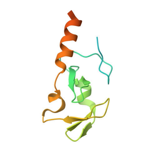
Structure and catalytic activation of the TRIM23 RING E3 ubiquitin ligase.

Dawidziak, D.M.Sanchez, J.G.Wagner, J.M.Ganser-Pornillos, B.K.Pornillos, O.

(2017) Proteins 85: 1957-1961

  • DOI: https://doi.org/10.1002/prot.25348
  • Primary Citation of Related Structures:  
    5VZV, 5VZW

  • PubMed Abstract: 

    Tripartite motif (TRIM) proteins comprise a large family of RING-type ubiquitin E3 ligases that regulate important biological processes. An emerging general model is that TRIMs form elongated antiparallel coiled-coil dimers that prevent interaction of the two attendant RING domains. The RING domains themselves bind E2 conjugating enzymes as dimers, implying that an active TRIM ligase requires higher-order oligomerization of the basal coiled-coil dimers. Here, we report crystal structures of the TRIM23 RING domain in isolation and in complex with an E2-ubiquitin conjugate. Our results indicate that TRIM23 enzymatic activity requires RING dimerization, consistent with the general model of TRIM activation.


  • Organizational Affiliation

    Department of Molecular Physiology and Biological Physics, University of Virginia, Charlottesville, Virginia.


Macromolecules
Find similar proteins by:  (by identity cutoff)  |  3D Structure
Entity ID: 1
MoleculeChains Sequence LengthOrganismDetailsImage
E3 ubiquitin-protein ligase TRIM23
A, B, C
123Homo sapiensMutation(s): 0 
Gene Names: TRIM23ARD1ARFD1RNF46
EC: 2.3.2.27
UniProt & NIH Common Fund Data Resources
Find proteins for P36406 (Homo sapiens)
Explore P36406 
Go to UniProtKB:  P36406
PHAROS:  P36406
GTEx:  ENSG00000113595 
Entity Groups  
Sequence Clusters30% Identity50% Identity70% Identity90% Identity95% Identity100% Identity
UniProt GroupP36406
Sequence Annotations
Expand
  • Reference Sequence
Experimental Data & Validation

Experimental Data

  • Method: X-RAY DIFFRACTION
  • Resolution: 1.81 Å
  • R-Value Free: 0.221 
  • R-Value Work: 0.186 
  • R-Value Observed: 0.189 
  • Space Group: C 2 2 21
Unit Cell:
Length ( Å )Angle ( ˚ )
a = 91.717α = 90
b = 142.464β = 90
c = 45.427γ = 90
Software Package:
Software NamePurpose
PHENIXrefinement
HKL-2000data reduction
HKL-2000data scaling
PHENIXphasing

Structure Validation

View Full Validation Report


Entry History & Funding Information

Deposition Data


Funding OrganizationLocationGrant Number
National Institutes of Health/National Institute of General Medical Sciences (NIH/NIGMS)United StatesR01-GM112508

Revision History  (Full details and data files)

  • Version 1.0: 2017-08-09
    Type: Initial release
  • Version 1.1: 2017-09-20
    Changes: Database references
  • Version 1.2: 2020-01-01
    Changes: Author supporting evidence
  • Version 1.3: 2024-03-13
    Changes: Data collection, Database references
  • Version 1.4: 2024-04-03
    Changes: Refinement description