5VPE

Transcription factor FosB/JunD bZIP domain in complex with cognate DNA, type-I crystal


Experimental Data Snapshot

  • Method: X-RAY DIFFRACTION
  • Resolution: 2.05 Å
  • R-Value Free: 0.254 
  • R-Value Work: 0.218 
  • R-Value Observed: 0.219 

wwPDB Validation   3D Report Full Report


This is version 1.4 of the entry. See complete history


Literature

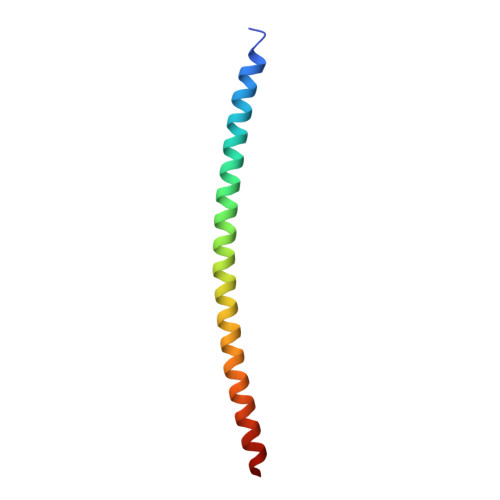
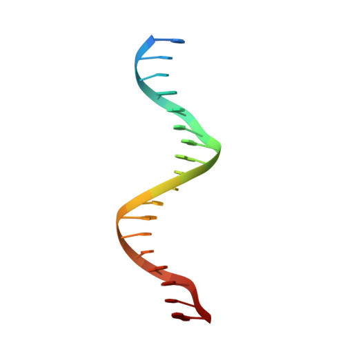
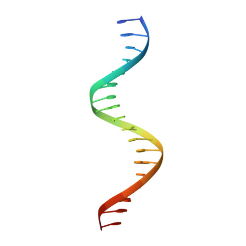
Activator Protein-1: redox switch controlling structure and DNA-binding.

Yin, Z.Machius, M.Nestler, E.J.Rudenko, G.

(2017) Nucleic Acids Res 45: 11425-11436

  • DOI: https://doi.org/10.1093/nar/gkx795
  • Primary Citation of Related Structures:  
    5VPA, 5VPB, 5VPC, 5VPD, 5VPE, 5VPF

  • PubMed Abstract: 

    The transcription factor, activator protein-1 (AP-1), binds to cognate DNA under redox control; yet, the underlying mechanism has remained enigmatic. A series of crystal structures of the AP-1 FosB/JunD bZIP domains reveal ordered DNA-binding regions in both FosB and JunD even in absence DNA. However, while JunD is competent to bind DNA, the FosB bZIP domain must undergo a large conformational rearrangement that is controlled by a 'redox switch' centered on an inter-molecular disulfide bond. Solution studies confirm that FosB/JunD cannot undergo structural transition and bind DNA when the redox-switch is in the 'OFF' state, and show that the mid-point redox potential of the redox switch affords it sensitivity to cellular redox homeostasis. The molecular and structural studies presented here thus reveal the mechanism underlying redox-regulation of AP-1 Fos/Jun transcription factors and provide structural insight for therapeutic interventions targeting AP-1 proteins.


  • Organizational Affiliation

    Department of Pharmacology and Toxicology, and the Sealy Center for Structural Biology, University of Texas Medical Branch, Galveston, TX 77555, USA.


Macromolecules

Find similar proteins by:  (by identity cutoff)  |  3D Structure
Entity ID: 1
MoleculeChains Sequence LengthOrganismDetailsImage
Protein fosBA [auth C],
E [auth A]
68Homo sapiensMutation(s): 0 
Gene Names: FOSBG0S3
UniProt & NIH Common Fund Data Resources
Find proteins for P53539 (Homo sapiens)
Explore P53539 
Go to UniProtKB:  P53539
PHAROS:  P53539
GTEx:  ENSG00000125740 
Entity Groups  
Sequence Clusters30% Identity50% Identity70% Identity90% Identity95% Identity100% Identity
UniProt GroupP53539
Sequence Annotations
Expand
  • Reference Sequence
Find similar proteins by:  (by identity cutoff)  |  3D Structure
Entity ID: 2
MoleculeChains Sequence LengthOrganismDetailsImage
Transcription factor jun-DB [auth D],
F [auth B]
68Homo sapiensMutation(s): 0 
Gene Names: JUND
UniProt & NIH Common Fund Data Resources
Find proteins for P17535 (Homo sapiens)
Explore P17535 
Go to UniProtKB:  P17535
PHAROS:  P17535
GTEx:  ENSG00000130522 
Entity Groups  
Sequence Clusters30% Identity50% Identity70% Identity90% Identity95% Identity100% Identity
UniProt GroupP17535
Sequence Annotations
Expand
  • Reference Sequence

Find similar nucleic acids by:  Sequence   |   3D Structure  

Entity ID: 3
MoleculeChains LengthOrganismImage
DNA (5'-D(*CP*GP*TP*CP*GP*GP*TP*GP*AP*CP*TP*CP*AP*CP*CP*GP*AP*CP*G)-3')C [auth G],
G [auth E]
19Homo sapiens
Sequence Annotations
Expand
  • Reference Sequence

Find similar nucleic acids by:  Sequence   |   3D Structure  

Entity ID: 4
MoleculeChains LengthOrganismImage
DNA (5'-D(*CP*GP*TP*CP*GP*GP*TP*GP*AP*GP*TP*CP*AP*CP*CP*GP*AP*CP*G)-3')D [auth H],
H [auth F]
19Homo sapiens
Sequence Annotations
Expand
  • Reference Sequence
Small Molecules
Ligands 4 Unique
IDChains Name / Formula / InChI Key2D Diagram3D Interactions
PO4
Query on PO4

Download Ideal Coordinates CCD File 
I [auth C],
L [auth D],
Z [auth F]
PHOSPHATE ION
O4 P
NBIIXXVUZAFLBC-UHFFFAOYSA-K
EDO
Query on EDO

Download Ideal Coordinates CCD File 
J [auth C]
M [auth D]
N [auth D]
P [auth G]
R [auth A]
J [auth C],
M [auth D],
N [auth D],
P [auth G],
R [auth A],
S [auth A],
T [auth A],
U [auth B],
V [auth B],
W [auth B],
X [auth B]
1,2-ETHANEDIOL
C2 H6 O2
LYCAIKOWRPUZTN-UHFFFAOYSA-N
CL
Query on CL

Download Ideal Coordinates CCD File 
O [auth D]CHLORIDE ION
Cl
VEXZGXHMUGYJMC-UHFFFAOYSA-M
NA
Query on NA

Download Ideal Coordinates CCD File 
K [auth C],
Q [auth G],
Y [auth E]
SODIUM ION
Na
FKNQFGJONOIPTF-UHFFFAOYSA-N
Experimental Data & Validation

Experimental Data

  • Method: X-RAY DIFFRACTION
  • Resolution: 2.05 Å
  • R-Value Free: 0.254 
  • R-Value Work: 0.218 
  • R-Value Observed: 0.219 
  • Space Group: C 1 2 1
Unit Cell:
Length ( Å )Angle ( ˚ )
a = 116.479α = 90
b = 90.755β = 117.93
c = 105.618γ = 90
Software Package:
Software NamePurpose
PHENIXrefinement
SCALEPACKdata scaling
PDB_EXTRACTdata extraction
DENZOdata reduction
PHASERphasing

Structure Validation

View Full Validation Report


Entry History & Funding Information

Deposition Data


Funding OrganizationLocationGrant Number
National Institutes of Health/National Institute on Drug Abuse (NIH/NIDA)United StatesR01 DA040621
Brain and Behavior Research FoundationUnited States--

Revision History  (Full details and data files)

  • Version 1.0: 2017-09-06
    Type: Initial release
  • Version 1.1: 2017-10-18
    Changes: Database references
  • Version 1.2: 2017-11-15
    Changes: Database references
  • Version 1.3: 2019-12-11
    Changes: Author supporting evidence
  • Version 1.4: 2023-10-04
    Changes: Data collection, Database references, Refinement description