5TKM

Crystal structure of human APOBEC3B N-terminal Domain


Experimental Data Snapshot

  • Method: X-RAY DIFFRACTION
  • Resolution: 1.90 Å
  • R-Value Free: 0.203 
  • R-Value Work: 0.188 
  • R-Value Observed: 0.189 

wwPDB Validation   3D Report Full Report


This is version 1.3 of the entry. See complete history


Literature

Structural determinants of APOBEC3B non-catalytic domain for molecular assembly and catalytic regulation.

Xiao, X.Yang, H.Arutiunian, V.Fang, Y.Besse, G.Morimoto, C.Zirkle, B.Chen, X.S.

(2017) Nucleic Acids Res 45: 7494-7506

  • DOI: https://doi.org/10.1093/nar/gkx362
  • Primary Citation of Related Structures:  
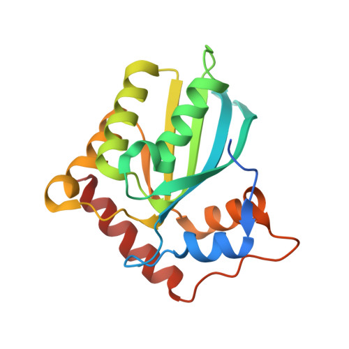
    5TKM

  • PubMed Abstract: 

    The catalytic activity of human cytidine deaminase APOBEC3B (A3B) has been correlated with kataegic mutational patterns within multiple cancer types. The molecular basis of how the N-terminal non-catalytic CD1 regulates the catalytic activity and consequently, biological function of A3B remains relatively unknown. Here, we report the crystal structure of a soluble human A3B-CD1 variant and delineate several structural elements of CD1 involved in molecular assembly, nucleic acid interactions and catalytic regulation of A3B. We show that (i) A3B expressed in human cells exists in hypoactive high-molecular-weight (HMW) complexes, which can be activated without apparent dissociation into low-molecular-weight (LMW) species after RNase A treatment. (ii) Multiple surface hydrophobic residues of CD1 mediate the HMW complex assembly and affect the catalytic activity, including one tryptophan residue W127 that likely acts through regulating nucleic acid binding. (iii) One of the highly positively charged surfaces on CD1 is involved in RNA-dependent attenuation of A3B catalysis. (iv) Surface hydrophobic residues of CD1 are involved in heterogeneous nuclear ribonucleoproteins (hnRNPs) binding to A3B. The structural and biochemical insights described here suggest that unique structural features on CD1 regulate the molecular assembly and catalytic activity of A3B through distinct mechanisms.


  • Organizational Affiliation

    Genetic, Molecular and Cellular Biology Program, Keck School of Medicine, University of Southern California, Los Angeles, CA 90089, USA.


Macromolecules
Find similar proteins by:  (by identity cutoff)  |  3D Structure
Entity ID: 1
MoleculeChains Sequence LengthOrganismDetailsImage
DNA dC->dU-editing enzyme APOBEC-3B
A, B
198Homo sapiensMutation(s): 7 
Gene Names: APOBEC3B
EC: 3.5.4
UniProt & NIH Common Fund Data Resources
Find proteins for Q9UH17 (Homo sapiens)
Explore Q9UH17 
Go to UniProtKB:  Q9UH17
PHAROS:  Q9UH17
GTEx:  ENSG00000179750 
Entity Groups  
Sequence Clusters30% Identity50% Identity70% Identity90% Identity95% Identity100% Identity
UniProt GroupQ9UH17
Sequence Annotations
Expand
  • Reference Sequence
Experimental Data & Validation

Experimental Data

  • Method: X-RAY DIFFRACTION
  • Resolution: 1.90 Å
  • R-Value Free: 0.203 
  • R-Value Work: 0.188 
  • R-Value Observed: 0.189 
  • Space Group: P 21 21 21
Unit Cell:
Length ( Å )Angle ( ˚ )
a = 60.739α = 90
b = 60.802β = 90
c = 111.515γ = 90
Software Package:
Software NamePurpose
PHENIXrefinement
HKL-2000data scaling
PHASERphasing

Structure Validation

View Full Validation Report


Entry History & Funding Information

Deposition Data


Funding OrganizationLocationGrant Number
National Institutes of Health/National Institute of General Medical Sciences (NIH/NIGMS)United StatesR01GM087986

Revision History  (Full details and data files)

  • Version 1.0: 2017-06-14
    Type: Initial release
  • Version 1.1: 2017-09-20
    Changes: Author supporting evidence, Database references, Refinement description
  • Version 1.2: 2019-12-25
    Changes: Author supporting evidence
  • Version 1.3: 2023-10-04
    Changes: Data collection, Database references, Derived calculations, Refinement description