5THV

Crystal Structure of G305A HDAC8 in complex with M344


Experimental Data Snapshot

  • Method: X-RAY DIFFRACTION
  • Resolution: 1.87 Å
  • R-Value Free: 0.210 
  • R-Value Work: 0.172 
  • R-Value Observed: 0.174 

wwPDB Validation   3D Report Full Report


Ligand Structure Quality Assessment 


This is version 1.4 of the entry. See complete history


Literature

Structural and Functional Influence of the Glycine-Rich Loop G302GGGY on the Catalytic Tyrosine of Histone Deacetylase 8.

Porter, N.J.Christianson, N.H.Decroos, C.Christianson, D.W.

(2016) Biochemistry 55: 6718-6729

  • DOI: https://doi.org/10.1021/acs.biochem.6b01014
  • Primary Citation of Related Structures:  
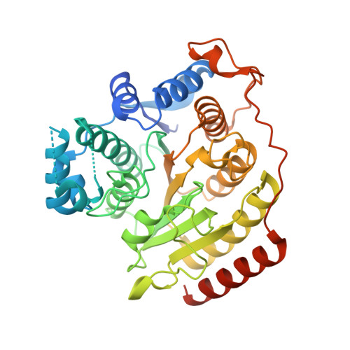
    5THS, 5THT, 5THU, 5THV

  • PubMed Abstract: 

    Histone deacetylase 8 (HDAC8) catalyzes the hydrolysis of acetyl-l-lysine to yield products l-lysine and acetate through a mechanism in which a nucleophilic water molecule is activated by a histidine general base and a catalytic metal ion (Zn 2+ or Fe 2+ ). Acetyl-l-lysine also requires activation by metal coordination and a hydrogen bond with catalytic tyrosine Y306, which also functions in transition state stabilization. Interestingly, Y306 is located in the conserved glycine-rich loop G 302 GGGY. The potential flexibility afforded by the tetraglycine segment may facilitate induced-fit conformational changes in Y306 between "in" and "out" positions, as observed in related deacetylases. To probe the catalytic importance of the glycine-rich loop in HDAC8, we rigidified this loop by preparing the G302A, G303A, G304A, and G305A mutants and measured their steady state kinetics and determined their X-ray crystal structures. Substantial losses of catalytic efficiency are observed (10-500-fold based on k cat /K M ), particularly for G304A HDAC8 and G305A HDAC8. These mutants also exhibit the greatest structural changes for catalytic tyrosine Y306 (1.3-1.7 Å shifts of the phenolic hydroxyl group). Molecular dynamics simulations further indicate that G304 and G305 undergo pronounced structural changes as residue 306 undergoes a transition between "in" and "out" conformations. Thus, the G304A and G305A substitutions likely compromise the position and conformational changes of Y306 required for substrate activation and transition state stabilization. The G302A and G303A substitutions have less severe catalytic consequences, and these substitutions may influence an internal channel through which product acetate is believed to exit.


  • Organizational Affiliation

    Roy and Diana Vagelos Laboratories, Department of Chemistry, University of Pennsylvania , Philadelphia, Pennsylvania 19104-6323, United States.


Macromolecules
Find similar proteins by:  (by identity cutoff)  |  3D Structure
Entity ID: 1
MoleculeChains Sequence LengthOrganismDetailsImage
Histone deacetylase 8
A, B
389Homo sapiensMutation(s): 1 
Gene Names: HDAC8HDACL1CDA07
EC: 3.5.1.98
UniProt & NIH Common Fund Data Resources
Find proteins for Q9BY41 (Homo sapiens)
Explore Q9BY41 
Go to UniProtKB:  Q9BY41
PHAROS:  Q9BY41
GTEx:  ENSG00000147099 
Entity Groups  
Sequence Clusters30% Identity50% Identity70% Identity90% Identity95% Identity100% Identity
UniProt GroupQ9BY41
Sequence Annotations
Expand
  • Reference Sequence
Small Molecules
Ligands 4 Unique
IDChains Name / Formula / InChI Key2D Diagram3D Interactions
B3N
Query on B3N

Download Ideal Coordinates CCD File 
F [auth A],
X [auth B]
4-(dimethylamino)-N-[7-(hydroxyamino)-7-oxoheptyl]benzamide
C16 H25 N3 O3
MXWDSZWTBOCWBK-UHFFFAOYSA-N
ZN
Query on ZN

Download Ideal Coordinates CCD File 
C [auth A],
U [auth B]
ZINC ION
Zn
PTFCDOFLOPIGGS-UHFFFAOYSA-N
EDO
Query on EDO

Download Ideal Coordinates CCD File 
AA [auth B]
BA [auth B]
CA [auth B]
DA [auth B]
EA [auth B]
AA [auth B],
BA [auth B],
CA [auth B],
DA [auth B],
EA [auth B],
FA [auth B],
G [auth A],
GA [auth B],
H [auth A],
HA [auth B],
I [auth A],
IA [auth B],
J [auth A],
JA [auth B],
K [auth A],
L [auth A],
M [auth A],
N [auth A],
O [auth A],
P [auth A],
Q [auth A],
R [auth A],
S [auth A],
T [auth A],
Y [auth B],
Z [auth B]
1,2-ETHANEDIOL
C2 H6 O2
LYCAIKOWRPUZTN-UHFFFAOYSA-N
K
Query on K

Download Ideal Coordinates CCD File 
D [auth A],
E [auth A],
V [auth B],
W [auth B]
POTASSIUM ION
K
NPYPAHLBTDXSSS-UHFFFAOYSA-N
Experimental Data & Validation

Experimental Data

  • Method: X-RAY DIFFRACTION
  • Resolution: 1.87 Å
  • R-Value Free: 0.210 
  • R-Value Work: 0.172 
  • R-Value Observed: 0.174 
  • Space Group: P 1 21 1
Unit Cell:
Length ( Å )Angle ( ˚ )
a = 53.03α = 90
b = 82.952β = 99.05
c = 94.55γ = 90
Software Package:
Software NamePurpose
PHENIXrefinement
iMOSFLMdata reduction
Aimlessdata scaling
PHASERphasing

Structure Validation

View Full Validation Report


Ligand Structure Quality Assessment 


Entry History & Funding Information

Deposition Data


Funding OrganizationLocationGrant Number
National Institutes of Health/National Institute of General Medical Sciences (NIH/NIGMS)United StatesGM49758

Revision History  (Full details and data files)

  • Version 1.0: 2016-12-21
    Type: Initial release
  • Version 1.1: 2017-09-27
    Changes: Author supporting evidence
  • Version 1.2: 2018-04-18
    Changes: Data collection, Database references, Derived calculations, Structure summary
  • Version 1.3: 2019-12-25
    Changes: Author supporting evidence, Derived calculations, Structure summary
  • Version 1.4: 2023-10-04
    Changes: Advisory, Data collection, Database references, Derived calculations, Refinement description