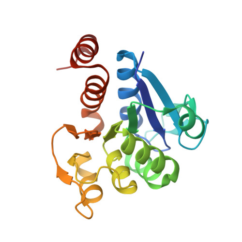
5SY6

Atomic resolution structure of human DJ-1, DTT bound


Experimental Data Snapshot

  • Method: X-RAY DIFFRACTION
  • Resolution: 1.15 Å
  • R-Value Free: 0.129 
  • R-Value Work: 0.110 
  • R-Value Observed: 0.111 

wwPDB Validation   3D Report Full Report


This is version 1.5 of the entry. See complete history


Literature

Short Carboxylic Acid-Carboxylate Hydrogen Bonds Can Have Fully Localized Protons.

Lin, J.Pozharski, E.Wilson, M.A.

(2017) Biochemistry 56: 391-402

  • DOI: https://doi.org/10.1021/acs.biochem.6b00906
  • Primary Citation of Related Structures:  
    5SY4, 5SY6, 5SY9, 5SYA

  • PubMed Abstract: 

    Short hydrogen bonds (H-bonds) have been proposed to play key functional roles in several proteins. The location of the proton in short H-bonds is of central importance, as proton delocalization is a defining feature of low-barrier hydrogen bonds (LBHBs). Experimentally determining proton location in H-bonds is challenging. Here, bond length analysis of atomic (1.15-0.98 Å) resolution X-ray crystal structures of the human protein DJ-1 and its bacterial homologue, YajL, was used to determine the protonation states of H-bonded carboxylic acids. DJ-1 contains a buried, dimer-spanning 2.49 Å H-bond between Glu15 and Asp24 that satisfies standard donor-acceptor distance criteria for a LBHB. Bond length analysis indicates that the proton is localized on Asp24, excluding a LBHB at this location. However, similar analysis of the Escherichia coli homologue YajL shows both residues may be protonated at the H-bonded oxygen atoms, potentially consistent with a LBHB. A Protein Data Bank-wide screen identifies candidate carboxylic acid H-bonds in approximately 14% of proteins, which are typically short [⟨d O-O ⟩ = 2.542(2) Å]. Chemically similar H-bonds between hydroxylated residues (Ser/Thr/Tyr) and carboxylates show a trend of lengthening O-O distance with increasing H-bond donor pK a . This trend suggests that conventional electronic effects provide an adequate explanation for short, charge-assisted carboxylic acid-carboxylate H-bonds in proteins, without the need to invoke LBHBs in general. This study demonstrates that bond length analysis of atomic resolution X-ray crystal structures provides a useful experimental test of certain candidate LBHBs.


  • Organizational Affiliation

    Department of Biochemistry and Redox Biology Center, University of Nebraska , Lincoln, Nebraska 68588, United States.


Macromolecules
Find similar proteins by:  (by identity cutoff)  |  3D Structure
Entity ID: 1
MoleculeChains Sequence LengthOrganismDetailsImage
Protein deglycase DJ-1192Homo sapiensMutation(s): 0 
Gene Names: PARK7
EC: 3.1.2 (PDB Primary Data), 3.5.1 (PDB Primary Data)
UniProt & NIH Common Fund Data Resources
Find proteins for Q99497 (Homo sapiens)
Explore Q99497 
Go to UniProtKB:  Q99497
PHAROS:  Q99497
GTEx:  ENSG00000116288 
Entity Groups  
Sequence Clusters30% Identity50% Identity70% Identity90% Identity95% Identity100% Identity
UniProt GroupQ99497
Sequence Annotations
Expand
  • Reference Sequence
Small Molecules
Ligands 1 Unique
IDChains Name / Formula / InChI Key2D Diagram3D Interactions
DTT
Query on DTT

Download Ideal Coordinates CCD File 
B [auth A]2,3-DIHYDROXY-1,4-DITHIOBUTANE
C4 H10 O2 S2
VHJLVAABSRFDPM-IMJSIDKUSA-N
Experimental Data & Validation

Experimental Data

  • Method: X-RAY DIFFRACTION
  • Resolution: 1.15 Å
  • R-Value Free: 0.129 
  • R-Value Work: 0.110 
  • R-Value Observed: 0.111 
  • Space Group: P 31 2 1
Unit Cell:
Length ( Å )Angle ( ˚ )
a = 75.227α = 90
b = 75.227β = 90
c = 75.227γ = 120
Software Package:
Software NamePurpose
REFMACrefinement
HKL-2000data reduction
Aimlessdata scaling
PHASERphasing

Structure Validation

View Full Validation Report


Entry History & Funding Information

Deposition Data


Funding OrganizationLocationGrant Number
National Institutes of Health/National Institute of General Medical Sciences (NIH/NIGMS)United StatesGM092999

Revision History  (Full details and data files)

  • Version 1.0: 2016-12-28
    Type: Initial release
  • Version 1.1: 2017-01-04
    Changes: Database references
  • Version 1.2: 2017-02-01
    Changes: Database references
  • Version 1.3: 2017-09-13
    Changes: Author supporting evidence
  • Version 1.4: 2019-12-25
    Changes: Author supporting evidence
  • Version 1.5: 2023-10-04
    Changes: Data collection, Database references, Refinement description