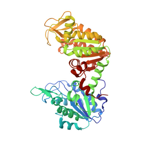
The phosphoglycerate kinase 1 variants found in carcinoma cells display different catalytic activity and conformational stability compared to the native enzyme.
Fiorillo, A., Petrosino, M., Ilari, A., Pasquo, A., Cipollone, A., Maggi, M., Chiaraluce, R., Consalvi, V.(2018) PLoS One 13: e0199191-e0199191
- PubMed: 29995887
- DOI: https://doi.org/10.1371/journal.pone.0199191
- Primary Citation of Related Structures:
5M1R, 5M3U, 5M6Z, 5MXM, 5O7D - PubMed Abstract:
Cancer cells are able to survive in difficult conditions, reprogramming their metabolism according to their requirements. Under hypoxic conditions they shift from oxidative phosphorylation to aerobic glycolysis, a behavior known as Warburg effect. In the last years, glycolytic enzymes have been identified as potential targets for alternative anticancer therapies. Recently, phosphoglycerate kinase 1 (PGK1), an ubiquitous enzyme expressed in all somatic cells that catalyzes the seventh step of glycolysis which consists of the reversible phosphotransfer reaction from 1,3-bisphosphoglycerate to ADP, has been discovered to be overexpressed in many cancer types. Moreover, several somatic variants of PGK1 have been identified in tumors. In this study we analyzed the effect of the single nucleotide variants found in cancer tissues on the PGK1 structure and function. Our results clearly show that the variants display a decreased catalytic efficiency and/or thermodynamic stability and an altered local tertiary structure, as shown by the solved X-ray structures. The changes in the catalytic properties and in the stability of the PGK1 variants, mainly due to the local changes evidenced by the X-ray structures, suggest also changes in the functional role of PGK to support the biosynthetic need of the growing and proliferating tumour cells.
Organizational Affiliation:
Department of Biochemical Sciences "A. Rossi Fanelli", Sapienza University of Rome, Rome, Italy.