5JMY

NEPRILYSIN COMPLEXED WITH LBQ657


Experimental Data Snapshot

  • Method: X-RAY DIFFRACTION
  • Resolution: 2.00 Å
  • R-Value Free: 0.228 
  • R-Value Work: 0.186 
  • R-Value Observed: 0.188 

wwPDB Validation   3D Report Full Report


Ligand Structure Quality Assessment 


This is version 2.1 of the entry. See complete history


Literature

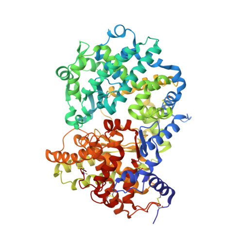
Structure of neprilysin in complex with the active metabolite of sacubitril.

Schiering, N.D'Arcy, A.Villard, F.Ramage, P.Logel, C.Cumin, F.Ksander, G.M.Wiesmann, C.Karki, R.G.Mogi, M.

(2016) Sci Rep 6: 27909-27909

  • DOI: https://doi.org/10.1038/srep27909
  • Primary Citation of Related Structures:  
    5JMY

  • PubMed Abstract: 

    Sacubitril is an ethyl ester prodrug of LBQ657, the active neprilysin (NEP) inhibitor, and a component of LCZ696 (sacubitril/valsartan). We report herein the three-dimensional structure of LBQ657 in complex with human NEP at 2 Å resolution. The crystal structure unravels the binding mode of the compound occupying the S1, S1' and S2' sub-pockets of the active site, consistent with a competitive inhibition mode. An induced fit conformational change upon binding of the P1'-biphenyl moiety of the inhibitor suggests an explanation for its selectivity against structurally homologous zinc metallopeptidases.


  • Organizational Affiliation

    Novartis Institutes for BioMedical Research Inc., Fabrikstrasse 16, CH-4002 Basel, Switzerland.


Macromolecules
Find similar proteins by:  (by identity cutoff)  |  3D Structure
Entity ID: 1
MoleculeChains Sequence LengthOrganismDetailsImage
Neprilysin
A, B
698Homo sapiensMutation(s): 0 
Gene Names: MMEEPN
EC: 3.4.24.11
UniProt & NIH Common Fund Data Resources
Find proteins for P08473 (Homo sapiens)
Explore P08473 
Go to UniProtKB:  P08473
PHAROS:  P08473
GTEx:  ENSG00000196549 
Entity Groups  
Sequence Clusters30% Identity50% Identity70% Identity90% Identity95% Identity100% Identity
UniProt GroupP08473
Sequence Annotations
Expand
  • Reference Sequence
Oligosaccharides

Help

Entity ID: 2
MoleculeChains Length2D Diagram Glycosylation3D Interactions
2-acetamido-2-deoxy-beta-D-glucopyranose-(1-4)-2-acetamido-2-deoxy-beta-D-glucopyranose
C, D
2N-Glycosylation
Glycosylation Resources
GlyTouCan:  G42666HT
GlyCosmos:  G42666HT
GlyGen:  G42666HT
Small Molecules
Ligands 3 Unique
IDChains Name / Formula / InChI Key2D Diagram3D Interactions
6LD
Query on 6LD

Download Ideal Coordinates CCD File 
I [auth A],
N [auth B]
Sacubitrilat
C22 H25 N O5
DOBNVUFHFMVMDB-BEFAXECRSA-N
NAG
Query on NAG

Download Ideal Coordinates CCD File 
E [auth A]
F [auth A]
G [auth A]
J [auth B]
K [auth B]
E [auth A],
F [auth A],
G [auth A],
J [auth B],
K [auth B],
L [auth B]
2-acetamido-2-deoxy-beta-D-glucopyranose
C8 H15 N O6
OVRNDRQMDRJTHS-FMDGEEDCSA-N
ZN
Query on ZN

Download Ideal Coordinates CCD File 
H [auth A],
M [auth B]
ZINC ION
Zn
PTFCDOFLOPIGGS-UHFFFAOYSA-N
Binding Affinity Annotations 
IDSourceBinding Affinity
6LD BindingDB:  5JMY IC50: min: 5, max: 20 (nM) from 4 assay(s)
Experimental Data & Validation

Experimental Data

  • Method: X-RAY DIFFRACTION
  • Resolution: 2.00 Å
  • R-Value Free: 0.228 
  • R-Value Work: 0.186 
  • R-Value Observed: 0.188 
  • Space Group: P 21 21 21
Unit Cell:
Length ( Å )Angle ( ˚ )
a = 59.737α = 90
b = 109.139β = 90
c = 248.003γ = 90
Software Package:
Software NamePurpose
XDSdata reduction
XSCALEdata scaling
PHASERphasing
BUSTERrefinement

Structure Validation

View Full Validation Report


Ligand Structure Quality Assessment 


Entry History 

Deposition Data

Revision History  (Full details and data files)

  • Version 1.0: 2016-06-15
    Type: Initial release
  • Version 1.1: 2016-06-29
    Changes: Database references
  • Version 1.2: 2019-04-24
    Changes: Data collection, Source and taxonomy
  • Version 2.0: 2020-07-29
    Type: Remediation
    Reason: Carbohydrate remediation
    Changes: Atomic model, Data collection, Derived calculations, Structure summary
  • Version 2.1: 2024-01-10
    Changes: Data collection, Database references, Refinement description, Structure summary