5I0I

Crystal structure of myosin X motor domain with 2IQ motifs in pre-powerstroke state


Experimental Data Snapshot

  • Method: X-RAY DIFFRACTION
  • Resolution: 3.15 Å
  • R-Value Free: 0.218 
  • R-Value Work: 0.192 
  • R-Value Observed: 0.193 

wwPDB Validation   3D Report Full Report


Ligand Structure Quality Assessment 


This is version 1.3 of the entry. See complete history


Literature

The myosin X motor is optimized for movement on actin bundles.

Ropars, V.Yang, Z.Isabet, T.Blanc, F.Zhou, K.Lin, T.Liu, X.Hissier, P.Samazan, F.Amigues, B.Yang, E.D.Park, H.Pylypenko, O.Cecchini, M.Sindelar, C.V.Sweeney, H.L.Houdusse, A.

(2016) Nat Commun 7: 12456-12456

  • DOI: https://doi.org/10.1038/ncomms12456
  • Primary Citation of Related Structures:  
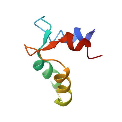
    5HMO, 5HMP, 5I0H, 5I0I, 5KG8

  • PubMed Abstract: 

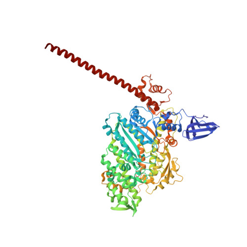
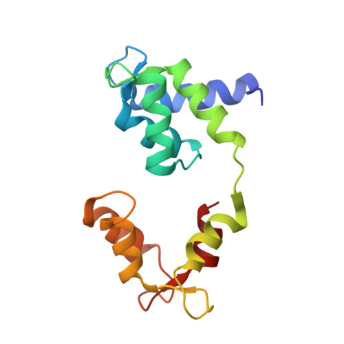
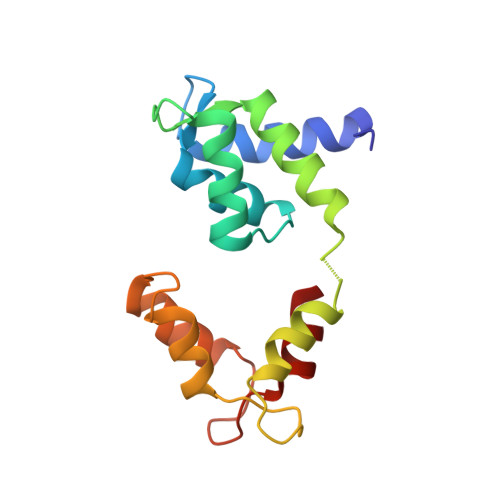
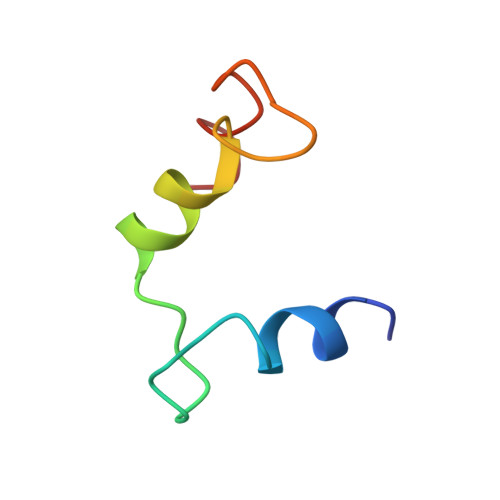
    Myosin X has features not found in other myosins. Its structure must underlie its unique ability to generate filopodia, which are essential for neuritogenesis, wound healing, cancer metastasis and some pathogenic infections. By determining high-resolution structures of key components of this motor, and characterizing the in vitro behaviour of the native dimer, we identify the features that explain the myosin X dimer behaviour. Single-molecule studies demonstrate that a native myosin X dimer moves on actin bundles with higher velocities and takes larger steps than on single actin filaments. The largest steps on actin bundles are larger than previously reported for artificially dimerized myosin X constructs or any other myosin. Our model and kinetic data explain why these large steps and high velocities can only occur on bundled filaments. Thus, myosin X functions as an antiparallel dimer in cells with a unique geometry optimized for movement on actin bundles.


  • Organizational Affiliation

    Structural Motility, Institut Curie, PSL Research University, CNRS, UMR 144, F-75005 Paris, France.


Macromolecules
Find similar proteins by:  (by identity cutoff)  |  3D Structure
Entity ID: 1
MoleculeChains Sequence LengthOrganismDetailsImage
Unconventional myosin-X
A, B
791Homo sapiensMutation(s): 0 
Gene Names: MYO10KIAA0799
UniProt & NIH Common Fund Data Resources
Find proteins for Q9HD67 (Homo sapiens)
Explore Q9HD67 
Go to UniProtKB:  Q9HD67
PHAROS:  Q9HD67
GTEx:  ENSG00000145555 
Entity Groups  
Sequence Clusters30% Identity50% Identity70% Identity90% Identity95% Identity100% Identity
UniProt GroupQ9HD67
Sequence Annotations
Expand
  • Reference Sequence
Find similar proteins by:  (by identity cutoff)  |  3D Structure
Entity ID: 2
MoleculeChains Sequence LengthOrganismDetailsImage
Calmodulin145Homo sapiensMutation(s): 0 
Gene Names: 
CALM1CALMCAMCAM1CALM2CAM2CAMBCALM3CALML2CAM3...
CALM1CALMCAMCAM1CALM2CAM2CAMBCALM3CALML2CAM3CAMCCAMIII

UniProt & NIH Common Fund Data Resources
Find proteins for P0DP23 (Homo sapiens)
Explore P0DP23 
Go to UniProtKB:  P0DP23
PHAROS:  P0DP23
GTEx:  ENSG00000198668 
Entity Groups  
Sequence Clusters30% Identity50% Identity70% Identity90% Identity95% Identity100% Identity
UniProt GroupP0DP23
Sequence Annotations
Expand
  • Reference Sequence
Find similar proteins by:  (by identity cutoff)  |  3D Structure
Entity ID: 3
MoleculeChains Sequence LengthOrganismDetailsImage
CalmodulinD [auth E]145Homo sapiensMutation(s): 0 
Gene Names: 
CALM1CALMCAMCAM1CALM2CAM2CAMBCALM3CALML2CAM3...
CALM1CALMCAMCAM1CALM2CAM2CAMBCALM3CALML2CAM3CAMCCAMIII

UniProt & NIH Common Fund Data Resources
Find proteins for P0DP23 (Homo sapiens)
Explore P0DP23 
Go to UniProtKB:  P0DP23
PHAROS:  P0DP23
GTEx:  ENSG00000198668 
Entity Groups  
Sequence Clusters30% Identity50% Identity70% Identity90% Identity95% Identity100% Identity
UniProt GroupP0DP23
Sequence Annotations
Expand
  • Reference Sequence
Find similar proteins by:  (by identity cutoff)  |  3D Structure
Entity ID: 4
MoleculeChains Sequence LengthOrganismDetailsImage
CalmodulinE [auth G]43Homo sapiensMutation(s): 0 
Gene Names: 
CALM1CALMCAMCAM1CALM2CAM2CAMBCALM3CALML2CAM3...
CALM1CALMCAMCAM1CALM2CAM2CAMBCALM3CALML2CAM3CAMCCAMIII

UniProt & NIH Common Fund Data Resources
Find proteins for P0DP23 (Homo sapiens)
Explore P0DP23 
Go to UniProtKB:  P0DP23
PHAROS:  P0DP23
GTEx:  ENSG00000198668 
Entity Groups  
Sequence Clusters30% Identity50% Identity70% Identity90% Identity95% Identity100% Identity
UniProt GroupP0DP23
Sequence Annotations
Expand
  • Reference Sequence
Find similar proteins by:  (by identity cutoff)  |  3D Structure
Entity ID: 5
MoleculeChains Sequence LengthOrganismDetailsImage
CalmodulinF [auth I]64Homo sapiensMutation(s): 0 
Gene Names: 
CALM1CALMCAMCAM1CALM2CAM2CAMBCALM3CALML2CAM3...
CALM1CALMCAMCAM1CALM2CAM2CAMBCALM3CALML2CAM3CAMCCAMIII

UniProt & NIH Common Fund Data Resources
Find proteins for P0DP23 (Homo sapiens)
Explore P0DP23 
Go to UniProtKB:  P0DP23
PHAROS:  P0DP23
GTEx:  ENSG00000198668 
Entity Groups  
Sequence Clusters30% Identity50% Identity70% Identity90% Identity95% Identity100% Identity
UniProt GroupP0DP23
Sequence Annotations
Expand
  • Reference Sequence
Small Molecules
Ligands 5 Unique
IDChains Name / Formula / InChI Key2D Diagram3D Interactions
ADP
Query on ADP

Download Ideal Coordinates CCD File 
J [auth A],
R [auth B]
ADENOSINE-5'-DIPHOSPHATE
C10 H15 N5 O10 P2
XTWYTFMLZFPYCI-KQYNXXCUSA-N
MPO
Query on MPO

Download Ideal Coordinates CCD File 
G [auth A],
P [auth B]
3[N-MORPHOLINO]PROPANE SULFONIC ACID
C7 H15 N O4 S
DVLFYONBTKHTER-UHFFFAOYSA-N
VO4
Query on VO4

Download Ideal Coordinates CCD File 
I [auth A],
S [auth B]
VANADATE ION
O4 V
LSGOVYNHVSXFFJ-UHFFFAOYSA-N
SO4
Query on SO4

Download Ideal Coordinates CCD File 
K [auth A]
L [auth A]
M [auth A]
N [auth A]
O [auth A]
K [auth A],
L [auth A],
M [auth A],
N [auth A],
O [auth A],
T [auth B],
U [auth C],
V [auth E]
SULFATE ION
O4 S
QAOWNCQODCNURD-UHFFFAOYSA-L
MG
Query on MG

Download Ideal Coordinates CCD File 
H [auth A],
Q [auth B]
MAGNESIUM ION
Mg
JLVVSXFLKOJNIY-UHFFFAOYSA-N
Experimental Data & Validation

Experimental Data

  • Method: X-RAY DIFFRACTION
  • Resolution: 3.15 Å
  • R-Value Free: 0.218 
  • R-Value Work: 0.192 
  • R-Value Observed: 0.193 
  • Space Group: P 21 21 21
Unit Cell:
Length ( Å )Angle ( ˚ )
a = 113.95α = 90
b = 173.41β = 90
c = 178.75γ = 90
Software Package:
Software NamePurpose
BUSTERrefinement
XDSdata reduction
PHASERphasing

Structure Validation

View Full Validation Report


Ligand Structure Quality Assessment 


Entry History & Funding Information

Deposition Data


Funding OrganizationLocationGrant Number
French National Research AgencyFranceANR blanche BLAN10
French National Research AgencyFranceANR-13-BSV8-0019-01
Ligue contre le cancerFrance--
ARCFrance--
National Institutes of HealthUnited StatesDC009100
National Institutes of HealthUnited StatesHL110869

Revision History  (Full details and data files)

  • Version 1.0: 2016-09-07
    Type: Initial release
  • Version 1.1: 2016-09-14
    Changes: Database references
  • Version 1.2: 2017-08-30
    Changes: Author supporting evidence
  • Version 1.3: 2017-11-22
    Changes: Database references