Structural complexity in the KCTD family of Cullin3-dependent E3 ubiquitin ligases.
Pinkas, D.M., Sanvitale, C.E., Bufton, J.C., Sorrell, F.J., Solcan, N., Chalk, R., Doutch, J., Bullock, A.N.(2017) Biochem J 474: 3747-3761
- PubMed: 28963344
- DOI: https://doi.org/10.1042/BCJ20170527
- Primary Citation of Related Structures:
4CRH, 4UIJ, 5A15, 5A6R, 5FTA - PubMed Abstract:
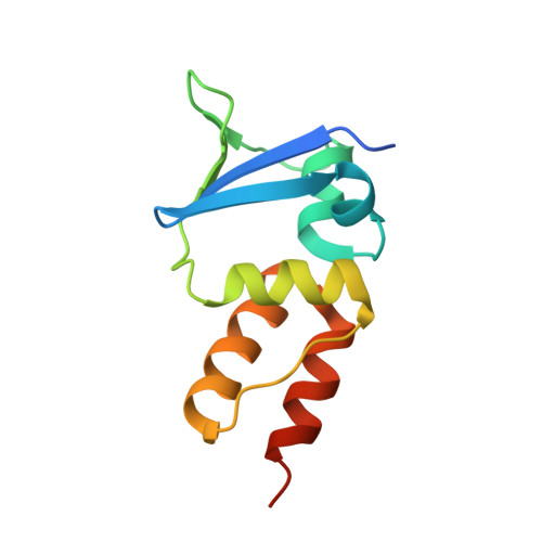
Members of the potassium channel tetramerization domain (KCTD) family are soluble non-channel proteins that commonly function as Cullin3 (Cul3)-dependent E3 ligases. Solution studies of the N-terminal BTB domain have suggested that some KCTD family members may tetramerize similarly to the homologous tetramerization domain (T1) of the voltage-gated potassium (Kv) channels. However, available structures of KCTD1, KCTD5 and KCTD9 have demonstrated instead pentameric assemblies. To explore other phylogenetic clades within the KCTD family, we determined the crystal structures of the BTB domains of a further five human KCTD proteins revealing a rich variety of oligomerization architectures, including monomer (SHKBP1), a novel two-fold symmetric tetramer (KCTD10 and KCTD13), open pentamer (KCTD16) and closed pentamer (KCTD17). While these diverse geometries were confirmed by small-angle X-ray scattering (SAXS), only the pentameric forms were stable upon size-exclusion chromatography. With the exception of KCTD16, all proteins bound to Cul3 and were observed to reassemble in solution as 5 : 5 heterodecamers. SAXS data and structural modelling indicate that Cul3 may stabilize closed BTB pentamers by binding across their BTB-BTB interfaces. These extra interactions likely also allow KCTD proteins to bind Cul3 without the expected 3-box motif. Overall, these studies reveal the KCTD family BTB domain to be a highly versatile scaffold compatible with a range of oligomeric assemblies and geometries. This observed interface plasticity may support functional changes in regulation of this unusual E3 ligase family.
Organizational Affiliation:
Structural Genomics Consortium, University of Oxford, Old Road Campus, Roosevelt Drive, Oxford OX3 7DQ, U.K.