5EHE

Crystal structure of human carbonic anhydrase isozyme II with 3-(benzylamino)-2,5,6-trifluoro-4-[(2-hydroxyethyl)sulfonyl]benzenesulfonamide


Experimental Data Snapshot

  • Method: X-RAY DIFFRACTION
  • Resolution: 1.50 Å
  • R-Value Free: 0.211 
  • R-Value Work: 0.180 
  • R-Value Observed: 0.183 

wwPDB Validation   3D Report Full Report


Ligand Structure Quality Assessment 


This is version 1.4 of the entry. See complete history


Literature

Intrinsic Thermodynamics and Structures of 2,4- and 3,4-Substituted Fluorinated Benzenesulfonamides Binding to Carbonic Anhydrases.

Zubriene, A.Smirnov, A.Dudutiene, V.Timm, D.D.Matuliene, J.Michailoviene, V.Zaksauskas, A.Manakova, E.Grazulis, S.Matulis, D.

(2017) ChemMedChem 12: 161-176

  • DOI: https://doi.org/10.1002/cmdc.201600509
  • Primary Citation of Related Structures:  
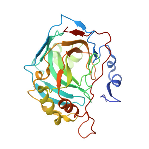
    5DOG, 5DOH, 5DRS, 5E2M, 5E2N, 5EHE

  • PubMed Abstract: 

    The goal of rational drug design is to understand structure-thermodynamics correlations in order to predict the chemical structure of a drug that would exhibit excellent affinity and selectivity for a target protein. In this study we explored the contribution of added functionalities of benzenesulfonamide inhibitors to the intrinsic binding affinity, enthalpy, and entropy for recombinant human carbonic anhydrases (CA) CA I, CA II, CA VII, CA IX, CA XII, and CA XIII. The binding enthalpies of compounds possessing similar chemical structures and affinities were found to be very different, spanning a range from -90 to +10 kJ mol -1 , and are compensated by a similar opposing entropy contribution. The intrinsic parameters of binding were determined by subtracting the linked protonation reactions. The sulfonamide group pK a values of the compounds were measured spectrophotometrically, and the protonation enthalpies were measured by isothermal titration calorimetry (ITC). Herein we describe the development of meta- or ortho-substituted fluorinated benzenesulfonamides toward the highly potent compound 10 h, which exhibits an observed dissociation constant value of 43 pm and an intrinsic dissociation constant value of 1.1 pm toward CA IX, an anticancer target that is highly overexpressed in various tumors. Fluorescence thermal shift assays, ITC, and X-ray crystallography were all applied in this work.


  • Organizational Affiliation

    Department of Biothermodynamics and Drug Design, Institute of Biotechnology, Life Sciences Center, Vilnius University, Saulėtekio 7, Vilnius, 10257, Lithuania.


Macromolecules
Find similar proteins by:  (by identity cutoff)  |  3D Structure
Entity ID: 1
MoleculeChains Sequence LengthOrganismDetailsImage
Carbonic anhydrase 2260Homo sapiensMutation(s): 0 
Gene Names: CA2
EC: 4.2.1.1
UniProt & NIH Common Fund Data Resources
Find proteins for P00918 (Homo sapiens)
Explore P00918 
Go to UniProtKB:  P00918
PHAROS:  P00918
GTEx:  ENSG00000104267 
Entity Groups  
Sequence Clusters30% Identity50% Identity70% Identity90% Identity95% Identity100% Identity
UniProt GroupP00918
Sequence Annotations
Expand
  • Reference Sequence
Small Molecules
Ligands 5 Unique
IDChains Name / Formula / InChI Key2D Diagram3D Interactions
WWO
Query on WWO

Download Ideal Coordinates CCD File 
G [auth A]3-(benzylamino)-2,5,6-trifluoro-4-[(2-hydroxyethyl)sulfonyl]benzenesulfonamide
C15 H15 F3 N2 O5 S2
OLTMJLATLWYQJF-UHFFFAOYSA-N
MES
Query on MES

Download Ideal Coordinates CCD File 
D [auth A]2-(N-MORPHOLINO)-ETHANESULFONIC ACID
C6 H13 N O4 S
SXGZJKUKBWWHRA-UHFFFAOYSA-N
PEG
Query on PEG

Download Ideal Coordinates CCD File 
E [auth A]DI(HYDROXYETHYL)ETHER
C4 H10 O3
MTHSVFCYNBDYFN-UHFFFAOYSA-N
DMS
Query on DMS

Download Ideal Coordinates CCD File 
C [auth A],
F [auth A]
DIMETHYL SULFOXIDE
C2 H6 O S
IAZDPXIOMUYVGZ-UHFFFAOYSA-N
ZN
Query on ZN

Download Ideal Coordinates CCD File 
B [auth A]ZINC ION
Zn
PTFCDOFLOPIGGS-UHFFFAOYSA-N
Binding Affinity Annotations 
IDSourceBinding Affinity
WWO BindingDB:  5EHE Kd: min: 0.04, max: 6.1 (nM) from 4 assay(s)
Experimental Data & Validation

Experimental Data

  • Method: X-RAY DIFFRACTION
  • Resolution: 1.50 Å
  • R-Value Free: 0.211 
  • R-Value Work: 0.180 
  • R-Value Observed: 0.183 
  • Space Group: P 1 21 1
Unit Cell:
Length ( Å )Angle ( ˚ )
a = 42.2α = 90
b = 41.026β = 104
c = 71.95γ = 90
Software Package:
Software NamePurpose
PDB_EXTRACTdata extraction
MOSFLMdata reduction
SCALAdata scaling
REFMACrefinement
MOLREPphasing
Cootmodel building

Structure Validation

View Full Validation Report


Ligand Structure Quality Assessment 


Entry History 

Deposition Data

Revision History  (Full details and data files)

  • Version 1.0: 2016-11-09
    Type: Initial release
  • Version 1.1: 2017-01-18
    Changes: Database references
  • Version 1.2: 2017-02-01
    Changes: Database references
  • Version 1.3: 2019-07-17
    Changes: Data collection
  • Version 1.4: 2024-01-10
    Changes: Data collection, Database references, Refinement description