5DU5

Crystal Structure of the ER-alpha Ligand-binding Domain in complex with a dichloro-substituted, 3,4-diarylthiophene dioxide core ligand


Experimental Data Snapshot

  • Method: X-RAY DIFFRACTION
  • Resolution: 2.19 Å
  • R-Value Free: 0.250 
  • R-Value Work: 0.199 
  • R-Value Observed: 0.203 

wwPDB Validation   3D Report Full Report


This is version 1.1 of the entry. See complete history


Literature

Predictive features of ligand-specific signaling through the estrogen receptor.

Nwachukwu, J.C.Srinivasan, S.Zheng, Y.Wang, S.Min, J.Dong, C.Liao, Z.Nowak, J.Wright, N.J.Houtman, R.Carlson, K.E.Josan, J.S.Elemento, O.Katzenellenbogen, J.A.Zhou, H.B.Nettles, K.W.

(2016) Mol Syst Biol 12: 864-864

  • DOI: https://doi.org/10.15252/msb.20156701
  • Primary Citation of Related Structures:  
    4ZN7, 4ZNH, 4ZNS, 4ZNT, 4ZNU, 4ZNV, 4ZNW, 5DI7, 5DID, 5DIE, 5DIG, 5DK9, 5DKB, 5DKE, 5DKG, 5DKS, 5DL4, 5DLR, 5DMC, 5DMF, 5DP0, 5DRJ, 5DRM, 5DTV, 5DU5, 5DUE, 5DUG, 5DUH, 5DVS, 5DVV, 5DWE, 5DWG, 5DWI, 5DWJ, 5DXK, 5DXM, 5DXP, 5DXQ, 5DXR, 5DY8, 5DYB, 5DYD, 5DZ0, 5DZ1, 5DZ3, 5DZH, 5DZI, 5E0W, 5E0X, 5E14

  • PubMed Abstract: 

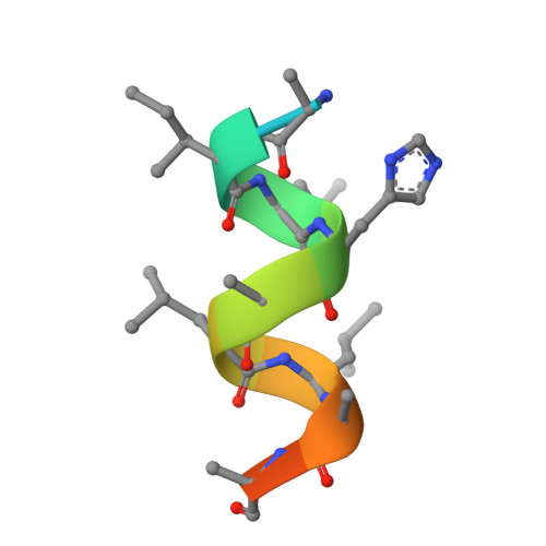
    Some estrogen receptor-α (ERα)-targeted breast cancer therapies such as tamoxifen have tissue-selective or cell-specific activities, while others have similar activities in different cell types. To identify biophysical determinants of cell-specific signaling and breast cancer cell proliferation, we synthesized 241 ERα ligands based on 19 chemical scaffolds, and compared ligand response using quantitative bioassays for canonical ERα activities and X-ray crystallography. Ligands that regulate the dynamics and stability of the coactivator-binding site in the C-terminal ligand-binding domain, called activation function-2 (AF-2), showed similar activity profiles in different cell types. Such ligands induced breast cancer cell proliferation in a manner that was predicted by the canonical recruitment of the coactivators NCOA1/2/3 and induction of the GREB1 proliferative gene. For some ligand series, a single inter-atomic distance in the ligand-binding domain predicted their proliferative effects. In contrast, the N-terminal coactivator-binding site, activation function-1 (AF-1), determined cell-specific signaling induced by ligands that used alternate mechanisms to control cell proliferation. Thus, incorporating systems structural analyses with quantitative chemical biology reveals how ligands can achieve distinct allosteric signaling outcomes through ERα.


  • Organizational Affiliation

    Department of Cancer Biology, The Scripps Research Institute, Jupiter, FL, USA.


Macromolecules
Find similar proteins by:  (by identity cutoff)  |  3D Structure
Entity ID: 1
MoleculeChains Sequence LengthOrganismDetailsImage
Estrogen receptor
A, B
257Homo sapiensMutation(s): 1 
Gene Names: ESR1ESRNR3A1
UniProt & NIH Common Fund Data Resources
Find proteins for P03372 (Homo sapiens)
Explore P03372 
Go to UniProtKB:  P03372
PHAROS:  P03372
GTEx:  ENSG00000091831 
Entity Groups  
Sequence Clusters30% Identity50% Identity70% Identity90% Identity95% Identity100% Identity
UniProt GroupP03372
Sequence Annotations
Expand
  • Reference Sequence

Find similar proteins by:  Sequence   |   3D Structure  

Entity ID: 2
MoleculeChains Sequence LengthOrganismDetailsImage
Nuclear receptor coactivator 2
C, D
14Homo sapiensMutation(s): 0 
UniProt & NIH Common Fund Data Resources
Find proteins for Q15596 (Homo sapiens)
Explore Q15596 
Go to UniProtKB:  Q15596
PHAROS:  Q15596
GTEx:  ENSG00000140396 
Entity Groups  
Sequence Clusters30% Identity50% Identity70% Identity90% Identity95% Identity100% Identity
UniProt GroupQ15596
Sequence Annotations
Expand
  • Reference Sequence
Small Molecules
Ligands 1 Unique
IDChains Name / Formula / InChI Key2D Diagram3D Interactions
5G2
Query on 5G2

Download Ideal Coordinates CCD File 
E [auth A],
F [auth B]
3,4-bis(2-chloro-4-hydroxyphenyl)-1H-1lambda~6~-thiophene-1,1-dione
C16 H10 Cl2 O4 S
YAJJGZGGQSGJNJ-UHFFFAOYSA-N
Experimental Data & Validation

Experimental Data

  • Method: X-RAY DIFFRACTION
  • Resolution: 2.19 Å
  • R-Value Free: 0.250 
  • R-Value Work: 0.199 
  • R-Value Observed: 0.203 
  • Space Group: P 1 21 1
Unit Cell:
Length ( Å )Angle ( ˚ )
a = 55.482α = 90
b = 82.712β = 110.75
c = 58.942γ = 90
Software Package:
Software NamePurpose
PHENIXrefinement
HKL-2000data reduction
HKL-2000data scaling
PHENIXphasing

Structure Validation

View Full Validation Report


Entry History 

Revision History  (Full details and data files)

  • Version 1.0: 2016-05-04
    Type: Initial release
  • Version 1.1: 2023-09-27
    Changes: Data collection, Database references, Derived calculations, Refinement description