Tetramerization and interdomain flexibility of the replication initiation controller YabA enables simultaneous binding to multiple partners.
Felicori, L., Jameson, K.H., Roblin, P., Fogg, M.J., Garcia-Garcia, T., Ventroux, M., Cherrier, M.V., Bazin, A., Noirot, P., Wilkinson, A.J., Molina, F., Terradot, L., Noirot-Gros, M.F.(2016) Nucleic Acids Res 44: 449-463
- PubMed: 26615189
- DOI: https://doi.org/10.1093/nar/gkv1318
- Primary Citation of Related Structures:
5DOL - PubMed Abstract:
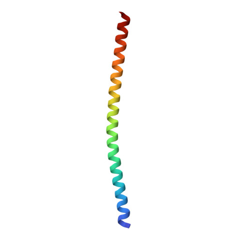
YabA negatively regulates initiation of DNA replication in low-GC Gram-positive bacteria. The protein exerts its control through interactions with the initiator protein DnaA and the sliding clamp DnaN. Here, we combined X-ray crystallography, X-ray scattering (SAXS), modeling and biophysical approaches, with in vivo experimental data to gain insight into YabA function. The crystal structure of the N-terminal domain (NTD) of YabA solved at 2.7 Å resolution reveals an extended α-helix that contributes to an intermolecular four-helix bundle. Homology modeling and biochemical analysis indicates that the C-terminal domain (CTD) of YabA is a small Zn-binding domain. Multi-angle light scattering and SAXS demonstrate that YabA is a tetramer in which the CTDs are independent and connected to the N-terminal four-helix bundle via flexible linkers. While YabA can simultaneously interact with both DnaA and DnaN, we found that an isolated CTD can bind to either DnaA or DnaN, individually. Site-directed mutagenesis and yeast-two hybrid assays identified DnaA and DnaN binding sites on the YabA CTD that partially overlap and point to a mutually exclusive mode of interaction. Our study defines YabA as a novel structural hub and explains how the protein tetramer uses independent CTDs to bind multiple partners to orchestrate replication initiation in the bacterial cell.
Organizational Affiliation:
Departamento de Bioquimica e Imunologia, Universidade Federal de Minas Gerais, UFMG, 31270-901, Belo Horizonte, MG, Brazil Sys2Diag FRE3690-CNRS/ALCEDIAG, Montpellier, France.


