5C23

Parkin (S65DUblR0RBR)


Experimental Data Snapshot

  • Method: X-RAY DIFFRACTION
  • Resolution: 2.37 Å
  • R-Value Free: 0.241 
  • R-Value Work: 0.220 
  • R-Value Observed: 0.221 

wwPDB Validation   3D Report Full Report


This is version 1.3 of the entry. See complete history


Literature

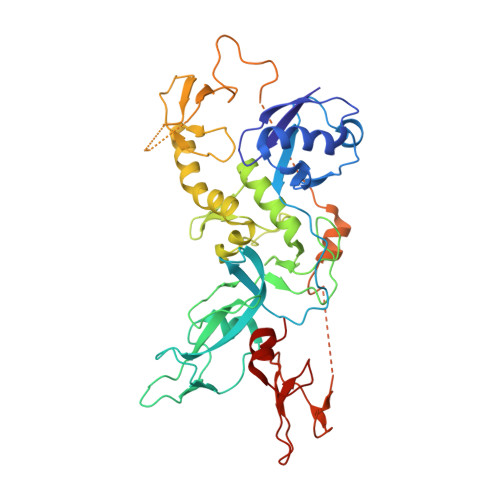
Disruption of the autoinhibited state primes the E3 ligase parkin for activation and catalysis.

Kumar, A.Aguirre, J.D.Condos, T.E.Martinez-Torres, R.J.Chaugule, V.K.Toth, R.Sundaramoorthy, R.Mercier, P.Knebel, A.Spratt, D.E.Barber, K.R.Shaw, G.S.Walden, H.

(2015) EMBO J 34: 2506-2521

  • DOI: https://doi.org/10.15252/embj.201592337
  • Primary Citation of Related Structures:  
    5C1Z, 5C23

  • PubMed Abstract: 

    The PARK2 gene is mutated in 50% of autosomal recessive juvenile parkinsonism (ARJP) cases. It encodes parkin, an E3 ubiquitin ligase of the RBR family. Parkin exists in an autoinhibited state that is activated by phosphorylation of its N-terminal ubiquitin-like (Ubl) domain and binding of phosphoubiquitin. We describe the 1.8 Å crystal structure of human parkin in its fully inhibited state and identify the key interfaces to maintain parkin inhibition. We identify the phosphoubiquitin-binding interface, provide a model for the phosphoubiquitin-parkin complex and show how phosphorylation of the Ubl domain primes parkin for optimal phosphoubiquitin binding. Furthermore, we demonstrate that the addition of phosphoubiquitin leads to displacement of the Ubl domain through loss of structure, unveiling a ubiquitin-binding site used by the E2~Ub conjugate, thus leading to active parkin. We find the role of the Ubl domain is to prevent parkin activity in the absence of the phosphorylation signals, and propose a model for parkin inhibition, optimization for phosphoubiquitin recruitment, release of inhibition by the Ubl domain and engagement with an E2~Ub conjugate. Taken together, this model provides a mechanistic framework for activating parkin.


  • Organizational Affiliation

    MRC Protein Phosphorylation and Ubiquitylation Unit, College of Life Sciences University of Dundee, Dundee, UK.


Macromolecules
Find similar proteins by:  (by identity cutoff)  |  3D Structure
Entity ID: 1
MoleculeChains Sequence LengthOrganismDetailsImage
E3 ubiquitin-protein ligase parkin
A, B
405Homo sapiensMutation(s): 0 
Gene Names: PARK2PRKN
EC: 6.3.2
UniProt & NIH Common Fund Data Resources
Find proteins for O60260 (Homo sapiens)
Explore O60260 
Go to UniProtKB:  O60260
PHAROS:  O60260
GTEx:  ENSG00000185345 
Entity Groups  
Sequence Clusters30% Identity50% Identity70% Identity90% Identity95% Identity100% Identity
UniProt GroupO60260
Sequence Annotations
Expand
  • Reference Sequence
Small Molecules
Ligands 4 Unique
IDChains Name / Formula / InChI Key2D Diagram3D Interactions
SO4
Query on SO4

Download Ideal Coordinates CCD File 
M [auth A],
N [auth A],
Z [auth B]
SULFATE ION
O4 S
QAOWNCQODCNURD-UHFFFAOYSA-L
GOL
Query on GOL

Download Ideal Coordinates CCD File 
K [auth A],
L [auth A],
X [auth B],
Y [auth B]
GLYCEROL
C3 H8 O3
PEDCQBHIVMGVHV-UHFFFAOYSA-N
ZN
Query on ZN

Download Ideal Coordinates CCD File 
C [auth A]
D [auth A]
E [auth A]
F [auth A]
G [auth A]
C [auth A],
D [auth A],
E [auth A],
F [auth A],
G [auth A],
H [auth A],
I [auth A],
J [auth A],
P [auth B],
Q [auth B],
R [auth B],
S [auth B],
T [auth B],
U [auth B],
V [auth B],
W [auth B]
ZINC ION
Zn
PTFCDOFLOPIGGS-UHFFFAOYSA-N
CL
Query on CL

Download Ideal Coordinates CCD File 
O [auth A]CHLORIDE ION
Cl
VEXZGXHMUGYJMC-UHFFFAOYSA-M
Experimental Data & Validation

Experimental Data

  • Method: X-RAY DIFFRACTION
  • Resolution: 2.37 Å
  • R-Value Free: 0.241 
  • R-Value Work: 0.220 
  • R-Value Observed: 0.221 
  • Space Group: P 21 21 21
Unit Cell:
Length ( Å )Angle ( ˚ )
a = 66.279α = 90
b = 66.29β = 90
c = 205.639γ = 90
Software Package:
Software NamePurpose
BUSTERrefinement
iMOSFLMdata reduction
SCALAdata scaling
BALBESphasing

Structure Validation

View Full Validation Report


Entry History 

Revision History  (Full details and data files)

  • Version 1.0: 2015-07-29
    Type: Initial release
  • Version 1.1: 2015-08-19
    Changes: Database references
  • Version 1.2: 2015-10-28
    Changes: Database references
  • Version 1.3: 2024-01-10
    Changes: Data collection, Database references, Refinement description