5AEA

Crystal structure of human NCAM domain 1


Experimental Data Snapshot

  • Method: X-RAY DIFFRACTION
  • Resolution: 1.90 Å
  • R-Value Free: 0.199 
  • R-Value Work: 0.172 
  • R-Value Observed: 0.174 

wwPDB Validation   3D Report Full Report


This is version 2.1 of the entry. See complete history


Literature

I-Bodies, Human Single Domain Antibodies that Antagonize Chemokine Receptor Cxcr4.

Griffiths, K.Dolezal, O.Cao, B.Nilsson, S.K.See, H.B.Pfleger, K.D.G.Roche, M.Gorry, P.R.Pow, A.Viduka, K.Lim, K.Lu, B.G.C.Chang, D.H.C.Murray-Rust, T.Kvansakul, M.Perugini, M.A.Dogovski, C.Doerflinger, M.Zhang, Y.Parisi, K.Casey, J.L.Nuttall, S.D.Foley, M.

(2016) J Biol Chem 291: 12641

  • DOI: https://doi.org/10.1074/jbc.M116.721050
  • Primary Citation of Related Structures:  
    5AEA

  • PubMed Abstract: 

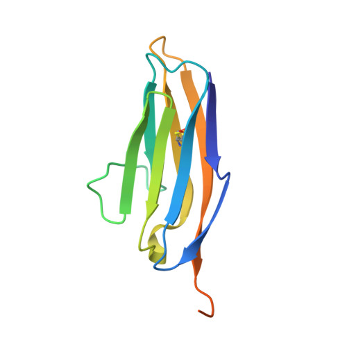
    CXCR4 is a G protein-coupled receptor with excellent potential as a therapeutic target for a range of clinical conditions, including stem cell mobilization, cancer prognosis and treatment, fibrosis therapy, and HIV infection. We report here the development of a fully human single-domain antibody-like scaffold termed an "i-body," the engineering of which produces an i-body library possessing a long complementarity determining region binding loop, and the isolation and characterization of a panel of i-bodies with activity against human CXCR4. The CXCR4-specific i-bodies show antagonistic activity in a range of in vitro and in vivo assays, including inhibition of HIV infection, cell migration, and leukocyte recruitment but, importantly, not the mobilization of hematopoietic stem cells. Epitope mapping of the three CXCR4 i-bodies AM3-114, AM4-272, and AM3-523 revealed binding deep in the binding pocket of the receptor.


  • Organizational Affiliation

    From AdAlta Pty. Ltd., 15/2 Park Dr., Bundoora, Victoria 3083.


Macromolecules
Find similar proteins by:  (by identity cutoff)  |  3D Structure
Entity ID: 1
MoleculeChains Sequence LengthOrganismDetailsImage
NEURAL CELL ADHESION MOLECULE 1
A, B
118Homo sapiensMutation(s): 0 
UniProt & NIH Common Fund Data Resources
Find proteins for P13591 (Homo sapiens)
Explore P13591 
Go to UniProtKB:  P13591
PHAROS:  P13591
GTEx:  ENSG00000149294 
Entity Groups  
Sequence Clusters30% Identity50% Identity70% Identity90% Identity95% Identity100% Identity
UniProt GroupP13591
Sequence Annotations
Expand
  • Reference Sequence
Small Molecules
Ligands 1 Unique
IDChains Name / Formula / InChI Key2D Diagram3D Interactions
FLC
Query on FLC

Download Ideal Coordinates CCD File 
C [auth B]CITRATE ANION
C6 H5 O7
KRKNYBCHXYNGOX-UHFFFAOYSA-K
Experimental Data & Validation

Experimental Data

  • Method: X-RAY DIFFRACTION
  • Resolution: 1.90 Å
  • R-Value Free: 0.199 
  • R-Value Work: 0.172 
  • R-Value Observed: 0.174 
  • Space Group: P 1 21 1
Unit Cell:
Length ( Å )Angle ( ˚ )
a = 23.85α = 90
b = 107.327β = 99.57
c = 41.748γ = 90
Software Package:
Software NamePurpose
PHENIXrefinement
XDSdata reduction
SCALAdata scaling
PHASERphasing

Structure Validation

View Full Validation Report


Entry History 

Deposition Data

Revision History  (Full details and data files)

  • Version 1.0: 2016-04-13
    Type: Initial release
  • Version 1.1: 2016-06-29
    Changes: Database references
  • Version 2.0: 2019-10-23
    Changes: Atomic model, Data collection, Other
  • Version 2.1: 2024-01-10
    Changes: Data collection, Database references, Derived calculations, Refinement description