5A3U

HIF prolyl hydroxylase 2 (PHD2/EGLN1) in complex with 6-(5-oxo-4-(1H- 1,2,3-triazol-1-yl)-2,5-dihydro-1H-pyrazol-1-yl)nicotinic acid


Experimental Data Snapshot

  • Method: X-RAY DIFFRACTION
  • Resolution: 3.30 Å
  • R-Value Free: 0.227 
  • R-Value Work: 0.202 
  • R-Value Observed: 0.192 

wwPDB Validation   3D Report Full Report


Ligand Structure Quality Assessment 


This is version 1.4 of the entry. See complete history


Literature

Potent and Selective Triazole-Based Inhibitors of the Hypoxia-Inducible Factor Prolyl-Hydroxylases with Activity in the Murine Brain.

Chan, M.C.Atasoylu, O.Hodson, E.Tumber, A.Leung, I.K.H.Chowdhury, R.Gomez-Perez, V.Demetriades, M.Rydzik, A.M.Holt-Martyn, J.Tian, Y.Bishop, T.Claridge, T.D.W.Kawamura, A.Pugh, C.W.Ratcliffe, P.J.Schofield, C.J.

(2015) PLoS One 10: 32004

  • DOI: https://doi.org/10.1371/journal.pone.0132004
  • Primary Citation of Related Structures:  
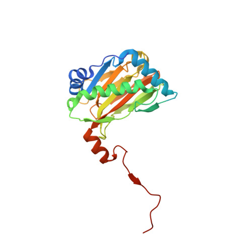
    5A3U

  • PubMed Abstract: 

    As part of the cellular adaptation to limiting oxygen availability in animals, the expression of a large set of genes is activated by the upregulation of the hypoxia-inducible transcription factors (HIFs). Therapeutic activation of the natural human hypoxic response can be achieved by the inhibition of the hypoxia sensors for the HIF system, i.e. the HIF prolyl-hydroxylases (PHDs). Here, we report studies on tricyclic triazole-containing compounds as potent and selective PHD inhibitors which compete with the 2-oxoglutarate co-substrate. One compound (IOX4) induces HIFα in cells and in wildtype mice with marked induction in the brain tissue, revealing that it is useful for studies aimed at validating the upregulation of HIF for treatment of cerebral diseases including stroke.


  • Organizational Affiliation

    Chemistry Research Laboratory, Department of Chemistry, University of Oxford, Oxford, United Kingdom; Centre for Cellular and Molecular Physiology, University of Oxford, Oxford, United Kingdom.


Macromolecules
Find similar proteins by:  (by identity cutoff)  |  3D Structure
Entity ID: 1
MoleculeChains Sequence LengthOrganismDetailsImage
EGL NINE HOMOLOG 1
A, B, C
252Homo sapiensMutation(s): 0 
EC: 1.14.11.2 (PDB Primary Data), 1.14.11.29 (PDB Primary Data), 1.14.11 (PDB Primary Data)
UniProt & NIH Common Fund Data Resources
Find proteins for Q9GZT9 (Homo sapiens)
Explore Q9GZT9 
Go to UniProtKB:  Q9GZT9
PHAROS:  Q9GZT9
GTEx:  ENSG00000135766 
Entity Groups  
Sequence Clusters30% Identity50% Identity70% Identity90% Identity95% Identity100% Identity
UniProt GroupQ9GZT9
Sequence Annotations
Expand
  • Reference Sequence
Experimental Data & Validation

Experimental Data

  • Method: X-RAY DIFFRACTION
  • Resolution: 3.30 Å
  • R-Value Free: 0.227 
  • R-Value Work: 0.202 
  • R-Value Observed: 0.192 
  • Space Group: P 32 1 2
Unit Cell:
Length ( Å )Angle ( ˚ )
a = 154.939α = 90
b = 154.939β = 90
c = 85.401γ = 120
Software Package:
Software NamePurpose
PHENIXrefinement
MOSFLMdata reduction
SCALAdata scaling
PHASERphasing

Structure Validation

View Full Validation Report


Ligand Structure Quality Assessment 


Entry History 

Deposition Data

Revision History  (Full details and data files)

  • Version 1.0: 2015-06-17
    Type: Initial release
  • Version 1.1: 2015-07-22
    Changes: Database references
  • Version 1.2: 2019-03-06
    Changes: Data collection, Experimental preparation, Other
  • Version 1.3: 2019-05-08
    Changes: Data collection, Experimental preparation
  • Version 1.4: 2024-01-10
    Changes: Data collection, Database references, Derived calculations, Other, Refinement description