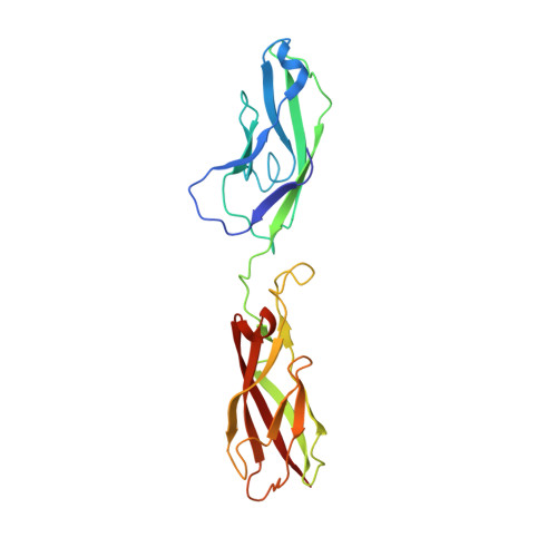
4ZTE

Crystal structure of human E-Cadherin (residues 3-213) in complex with a peptidomimetic inhibitor


Experimental Data Snapshot

  • Method: X-RAY DIFFRACTION
  • Resolution: 2.13 Å
  • R-Value Free: 0.230 
  • R-Value Work: 0.185 
  • R-Value Observed: 0.187 

wwPDB Validation   3D Report Full Report


Ligand Structure Quality Assessment 


This is version 1.2 of the entry. See complete history


Literature

Crystal Structure of Human E-Cadherin-EC1EC2 in Complex with a Peptidomimetic Competitive Inhibitor of Cadherin Homophilic Interaction.

Nardone, V.Lucarelli, A.P.Dalle Vedove, A.Fanelli, R.Tomassetti, A.Belvisi, L.Civera, M.Parisini, E.

(2016) J Med Chem 59: 5089-5094

  • DOI: https://doi.org/10.1021/acs.jmedchem.5b01487
  • Primary Citation of Related Structures:  
    4ZT1, 4ZTE

  • PubMed Abstract: 

    Cadherins are transmembrane cell adhesion proteins whose aberrant expression often correlates with cancer development and proliferation. We report the crystal structure of an E-cadherin extracellular fragment in complex with a peptidomimetic compound that was previously shown to partially inhibit cadherin homophilic adhesion. The structure reveals an unexpected binding mode and allows the identification of a druggable cadherin interface, thus paving the way to a future structure-guided design of cell adhesion inhibitors against cadherin-expressing solid tumors.


  • Organizational Affiliation

    Center for Nano Science and Technology @PoliMi, Istituto Italiano di Tecnologia , Via G. Pascoli 70/3, 20133 Milano, Italy.


Macromolecules
Find similar proteins by:  (by identity cutoff)  |  3D Structure
Entity ID: 1
MoleculeChains Sequence LengthOrganismDetailsImage
Cadherin-1
A, B
211Homo sapiensMutation(s): 0 
Gene Names: CDH1CDHEUVO
UniProt & NIH Common Fund Data Resources
Find proteins for P12830 (Homo sapiens)
Explore P12830 
Go to UniProtKB:  P12830
PHAROS:  P12830
GTEx:  ENSG00000039068 
Entity Groups  
Sequence Clusters30% Identity50% Identity70% Identity90% Identity95% Identity100% Identity
UniProt GroupP12830
Sequence Annotations
Expand
  • Reference Sequence
Small Molecules
Ligands 2 Unique
IDChains Name / Formula / InChI Key2D Diagram3D Interactions
4RL
Query on 4RL

Download Ideal Coordinates CCD File 
F [auth A],
J [auth B]
N-{[(2S,5S)-1-benzyl-5-(2-{[(2S,3S)-1-(tert-butylamino)-3-methyl-1-oxopentan-2-yl]amino}-2-oxoethyl)-3,6-dioxopiperazin-2-yl]methyl}-L-alpha-asparagine
C28 H42 N6 O7
QOSIRQNYWJNZFB-WGLQCKHLSA-N
CA
Query on CA

Download Ideal Coordinates CCD File 
C [auth A]
D [auth A]
E [auth A]
G [auth B]
H [auth B]
C [auth A],
D [auth A],
E [auth A],
G [auth B],
H [auth B],
I [auth B]
CALCIUM ION
Ca
BHPQYMZQTOCNFJ-UHFFFAOYSA-N
Experimental Data & Validation

Experimental Data

  • Method: X-RAY DIFFRACTION
  • Resolution: 2.13 Å
  • R-Value Free: 0.230 
  • R-Value Work: 0.185 
  • R-Value Observed: 0.187 
  • Space Group: C 1 2 1
Unit Cell:
Length ( Å )Angle ( ˚ )
a = 120.579α = 90
b = 76.835β = 111.14
c = 72.772γ = 90
Software Package:
Software NamePurpose
REFMACrefinement
SCALAdata scaling
MOLREPphasing
XDSdata reduction

Structure Validation

View Full Validation Report


Ligand Structure Quality Assessment 


Entry History & Funding Information

Deposition Data


Funding OrganizationLocationGrant Number
European UnionFP7-PEOPLE-2010-RG (N. 268231)

Revision History  (Full details and data files)

  • Version 1.0: 2016-06-01
    Type: Initial release
  • Version 1.1: 2016-06-08
    Changes: Database references
  • Version 1.2: 2024-01-10
    Changes: Data collection, Database references, Derived calculations, Refinement description