4XYN

X-ray structure of Ca(2+)-S100B with human RAGE-derived W61 peptide


Experimental Data Snapshot

  • Method: X-RAY DIFFRACTION
  • Resolution: 2.55 Å
  • R-Value Free: 0.254 
  • R-Value Work: 0.203 
  • R-Value Observed: 0.205 

wwPDB Validation   3D Report Full Report


This is version 1.4 of the entry. See complete history


Literature

Structural insights into the binding of the human receptor for advanced glycation end products (RAGE) by S100B, as revealed by an S100B-RAGE-derived peptide complex.

Jensen, J.L.Indurthi, V.S.Neau, D.B.Vetter, S.W.Colbert, C.L.

(2015) Acta Crystallogr D Biol Crystallogr 71: 1176-1183

  • DOI: https://doi.org/10.1107/S1399004715004216
  • Primary Citation of Related Structures:  
    4XYN

  • PubMed Abstract: 

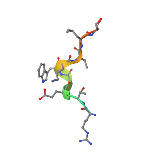
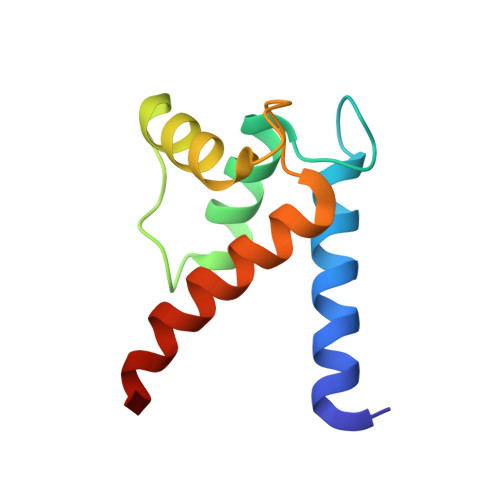
    S100B is a damage-associated molecular pattern protein that, when released into the extracellular milieu, triggers initiation of the inflammatory response through the receptor for advanced glycation end products (RAGE). Recognition of S100B is accomplished via the amino-terminal variable immunoglobulin domain (V-domain) of RAGE. To gain insights into this interaction, a complex between S100B and a 15-amino-acid peptide derived from residues 54-68 of the V-domain was crystallized. The X-ray crystal structure was solved to 2.55 Å resolution. There are two dimers of S100B and one peptide in the asymmetric unit. The binding interface of this peptide is compared with that found in the complex between S100B and the 12-amino-acid CapZ-derived peptide TRTK-12. This comparison reveals that although the peptides adopt completely different backbone structures, the residues buried at the interface interact with S100B in similar regions to form stable complexes. The binding affinities of S100B for the intact wild-type V-domain and a W61A V-domain mutant were determined to be 2.7 ± 0.5 and 1.3 ± 0.7 µM, respectively, using fluorescence titration experiments. These observations lead to a model whereby conformational flexibility in the RAGE receptor allows the adoption of a binding conformation for interaction with the stable hydrophobic groove on the surface of S100B.


  • Organizational Affiliation

    Department of Chemistry and Biochemistry, North Dakota State University, PO Box 6050, Fargo, ND 58108-6050, USA.


Macromolecules

Find similar proteins by:  Sequence   |   3D Structure  

Entity ID: 1
MoleculeChains Sequence LengthOrganismDetailsImage
Receptor for advanced glycation endproducts-derived peptide (W61)A [auth P]15synthetic constructMutation(s): 0 
UniProt & NIH Common Fund Data Resources
Find proteins for Q15109 (Homo sapiens)
Explore Q15109 
Go to UniProtKB:  Q15109
PHAROS:  Q15109
GTEx:  ENSG00000204305 
Entity Groups  
Sequence Clusters30% Identity50% Identity70% Identity90% Identity95% Identity100% Identity
UniProt GroupQ15109
Sequence Annotations
Expand
  • Reference Sequence
Find similar proteins by:  (by identity cutoff)  |  3D Structure
Entity ID: 2
MoleculeChains Sequence LengthOrganismDetailsImage
Protein S100-BB [auth A],
C [auth B],
D [auth C],
E [auth D]
92Homo sapiensMutation(s): 0 
Gene Names: S100B
UniProt & NIH Common Fund Data Resources
Find proteins for P04271 (Homo sapiens)
Explore P04271 
Go to UniProtKB:  P04271
PHAROS:  P04271
GTEx:  ENSG00000160307 
Entity Groups  
Sequence Clusters30% Identity50% Identity70% Identity90% Identity95% Identity100% Identity
UniProt GroupP04271
Sequence Annotations
Expand
  • Reference Sequence
Experimental Data & Validation

Experimental Data

  • Method: X-RAY DIFFRACTION
  • Resolution: 2.55 Å
  • R-Value Free: 0.254 
  • R-Value Work: 0.203 
  • R-Value Observed: 0.205 
  • Space Group: P 21 21 2
Unit Cell:
Length ( Å )Angle ( ˚ )
a = 68.785α = 90
b = 86.684β = 90
c = 66.534γ = 90
Software Package:
Software NamePurpose
PHENIXrefinement
ADSCdata collection
XDSdata scaling
autoPROCdata scaling
Aimlessdata scaling
Cootmodel building
BALBESphasing

Structure Validation

View Full Validation Report


Entry History 

Deposition Data

Revision History  (Full details and data files)

  • Version 1.0: 2015-05-13
    Type: Initial release
  • Version 1.1: 2015-05-20
    Changes: Database references
  • Version 1.2: 2015-05-27
    Changes: Data collection
  • Version 1.3: 2017-11-22
    Changes: Advisory, Derived calculations, Refinement description, Source and taxonomy
  • Version 1.4: 2024-02-28
    Changes: Advisory, Data collection, Database references, Derived calculations, Refinement description