4XH2

Crystal structure of human paxillin LD4 motif in complex with Fab fragment


Experimental Data Snapshot

  • Method: X-RAY DIFFRACTION
  • Resolution: 2.00 Å
  • R-Value Free: 0.258 
  • R-Value Work: 0.224 
  • R-Value Observed: 0.226 

wwPDB Validation   3D Report Full Report


Ligand Structure Quality Assessment 


This is version 1.4 of the entry. See complete history


Literature

Engineering Synthetic Antibody Inhibitors Specific for LD2 or LD4 Motifs of Paxillin.

Nocula-Lugowska, M.Lugowski, M.Salgia, R.Kossiakoff, A.A.

(2015) J Mol Biol 427: 2532-2547

  • DOI: https://doi.org/10.1016/j.jmb.2015.06.004
  • Primary Citation of Related Structures:  
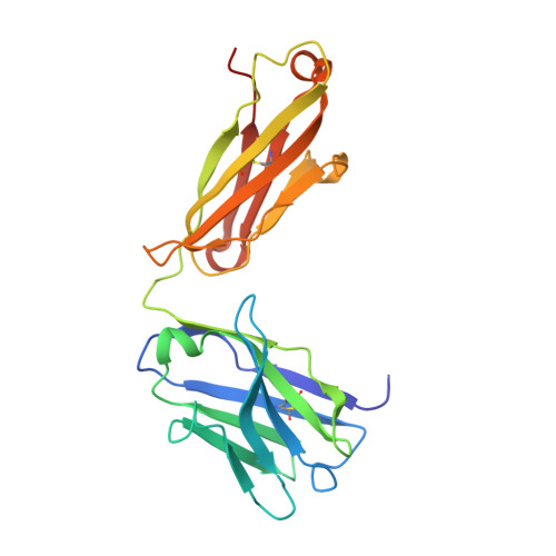
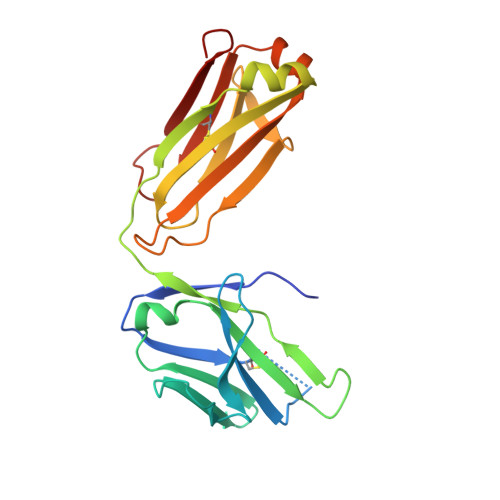
    4XGZ, 4XH2

  • PubMed Abstract: 

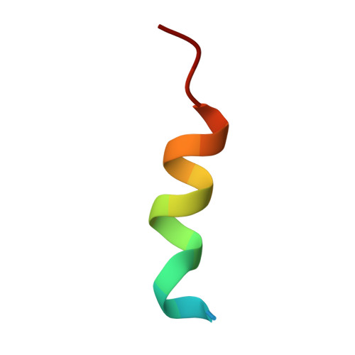
    Focal adhesion protein paxillin links integrin and growth factor signaling to actin cytoskeleton. Most of paxillin signaling activity is regulated via leucine-rich LD motifs (LD1-LD5) located at the N-terminus. Here, we demonstrate a method to engineer highly selective synthetic antibodies (sABs) against LD2 and LD4 that are binding sites for focal adhesion kinase (FAK) and other proteins. Phage display selections against peptides were used to generate sABs recognizing each LD motif. In the obtained X-ray crystal structures of the LD-sAB complexes, the LD motifs are helical and bind sABs through a hydrophobic side, similarly as in the structures with natural paxillin partners. The sABs are capable of pulling down endogenous paxillin in complex with FAK and can visualize paxillin in focal adhesions in cells. They were also used as selective inhibitors to effectively compete with focal adhesion targeting domain of FAK for the binding to LD2 and LD4. The sABs are tools for investigation of paxillin LD binding "platforms" and are capable of inhibiting paxillin interactions, thereby useful as potential therapeutics in the future.


  • Organizational Affiliation

    Department of Biochemistry and Molecular Biology, The University of Chicago, Chicago, IL 60607, USA.


Macromolecules
Find similar proteins by:  (by identity cutoff)  |  3D Structure
Entity ID: 1
MoleculeChains Sequence LengthOrganismDetailsImage
Fab Heavy Chain
A, C, E, G, H
A, C, E, G, H, J
228Homo sapiensMutation(s): 0 
Entity Groups  
Sequence Clusters30% Identity50% Identity70% Identity90% Identity95% Identity100% Identity
Sequence Annotations
Expand
  • Reference Sequence
Find similar proteins by:  (by identity cutoff)  |  3D Structure
Entity ID: 2
MoleculeChains Sequence LengthOrganismDetailsImage
Fab Light Chain
B, D, F, I, K
B, D, F, I, K, L
217Homo sapiensMutation(s): 0 
Entity Groups  
Sequence Clusters30% Identity50% Identity70% Identity90% Identity95% Identity100% Identity
Sequence Annotations
Expand
  • Reference Sequence

Find similar proteins by:  Sequence   |   3D Structure  

Entity ID: 3
MoleculeChains Sequence LengthOrganismDetailsImage
paxillin LD418synthetic constructMutation(s): 0 
UniProt & NIH Common Fund Data Resources
Find proteins for P49023 (Homo sapiens)
Explore P49023 
Go to UniProtKB:  P49023
PHAROS:  P49023
GTEx:  ENSG00000089159 
Entity Groups  
Sequence Clusters30% Identity50% Identity70% Identity90% Identity95% Identity100% Identity
UniProt GroupP49023
Sequence Annotations
Expand
  • Reference Sequence
Small Molecules
Ligands 5 Unique
IDChains Name / Formula / InChI Key2D Diagram3D Interactions
LDA
Query on LDA

Download Ideal Coordinates CCD File 
DA [auth F]LAURYL DIMETHYLAMINE-N-OXIDE
C14 H31 N O
SYELZBGXAIXKHU-UHFFFAOYSA-N
PO4
Query on PO4

Download Ideal Coordinates CCD File 
BA [auth F],
X [auth D]
PHOSPHATE ION
O4 P
NBIIXXVUZAFLBC-UHFFFAOYSA-K
GOL
Query on GOL

Download Ideal Coordinates CCD File 
AA [auth F]
EA [auth F]
FA [auth G]
GA [auth H]
HA [auth H]
AA [auth F],
EA [auth F],
FA [auth G],
GA [auth H],
HA [auth H],
IA [auth I],
JA [auth I],
KA [auth I],
MA [auth K],
NA [auth K],
OA [auth K],
PA [auth L],
QA [auth L],
S [auth B],
T [auth B],
U [auth C],
V [auth C],
W [auth D],
Y [auth F],
Z [auth F]
GLYCEROL
C3 H8 O3
PEDCQBHIVMGVHV-UHFFFAOYSA-N
ACT
Query on ACT

Download Ideal Coordinates CCD File 
CA [auth F]ACETATE ION
C2 H3 O2
QTBSBXVTEAMEQO-UHFFFAOYSA-M
ACE
Query on ACE

Download Ideal Coordinates CCD File 
LA [auth J],
RA [auth L]
ACETYL GROUP
C2 H4 O
IKHGUXGNUITLKF-UHFFFAOYSA-N
Experimental Data & Validation

Experimental Data

  • Method: X-RAY DIFFRACTION
  • Resolution: 2.00 Å
  • R-Value Free: 0.258 
  • R-Value Work: 0.224 
  • R-Value Observed: 0.226 
  • Space Group: P 21 21 21
Unit Cell:
Length ( Å )Angle ( ˚ )
a = 117.58α = 90
b = 138.12β = 90
c = 223.51γ = 90
Software Package:
Software NamePurpose
REFMACrefinement
XDSdata reduction
PDB_EXTRACTdata extraction
XSCALEdata scaling
PHASERphasing

Structure Validation

View Full Validation Report


Ligand Structure Quality Assessment 


Entry History & Funding Information

Deposition Data


Funding OrganizationLocationGrant Number
National Institutes of Health/National Institute of General Medical Sciences (NIH/NIGMS)United States--

Revision History  (Full details and data files)

  • Version 1.0: 2015-07-01
    Type: Initial release
  • Version 1.1: 2015-08-05
    Changes: Database references
  • Version 1.2: 2017-05-31
    Changes: Other
  • Version 1.3: 2019-11-27
    Changes: Author supporting evidence
  • Version 1.4: 2023-09-27
    Changes: Data collection, Database references, Refinement description