Aspartoacylase catalytic deficiency as the cause of canavan disease: a structural perspective.
Wijayasinghe, Y.S., Pavlovsky, A.G., Viola, R.E.(2014) Biochemistry 53: 4970-4978
- PubMed: 25003821
- DOI: https://doi.org/10.1021/bi500719k
- Primary Citation of Related Structures:
4MRI, 4MXU, 4NFR, 4TNU - PubMed Abstract:
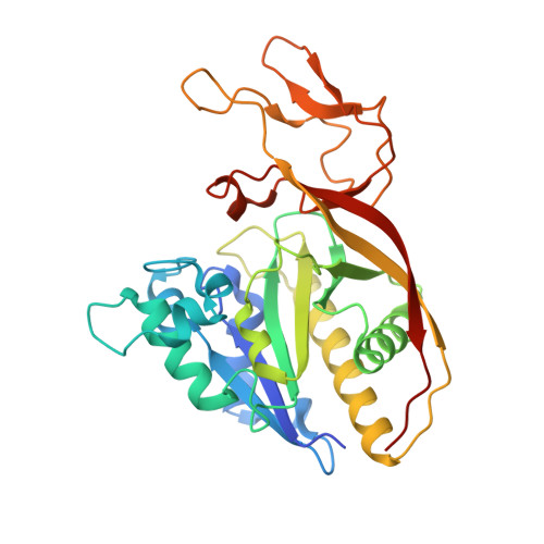
Canavan disease (CD) is a fatal, childhood neurological disorder caused by mutations in the ASPA gene, leading to catalytic deficiencies in the aspartoacylase (ASPA) enzyme and impaired N-acetyl-l-aspartic acid metabolism in the brain. To study the possible structural defects triggered by these mutations, four ASPA missense mutations associated with different disease severities have been structurally characterized. These mutant enzymes each have overall structures similar to that of the native ASPA enzyme, but with varying degrees of alterations that offer explanations for the respective loss of catalytic activity. The K213E mutant, a nonconservative mutant associated with a mild disease phenotype, has minimal structural differences compared to the native enzyme. In contrast, the loss of van der Waals contacts in the F295S mutant and the loss of hydrophobic and hydrogen bonding interactions in the Y231C mutant lead to a local collapse of the hydrophobic core structure in the carboxyl-terminal domain, contributing to a decrease in protein stability. The structure of the E285A mutant, the most common clinical mutant, reveals that the loss of hydrogen bonding interactions with the carboxylate side chain of Glu285 disturbs the active site architecture, leading to altered substrate binding and lower catalytic activity. Our improved understanding of the nature of these structural defects provides a basis for the development of treatment therapies for CD.
Organizational Affiliation:
Department of Chemistry and Biochemistry, The University of Toledo , Toledo, Ohio 43606, United States.