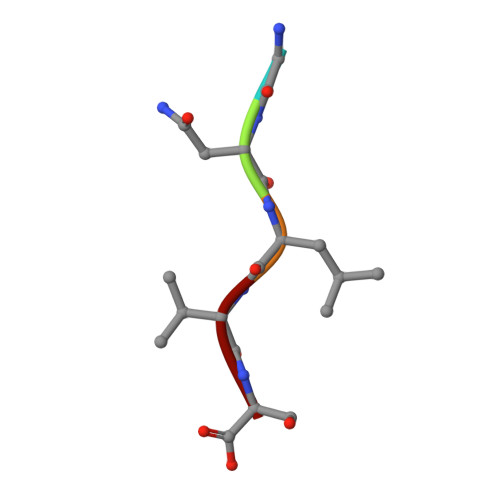
Toxicity of Eosinophil MBP Is Repressed by Intracellular Crystallization and Promoted by Extracellular Aggregation.
Soragni, A., Yousefi, S., Stoeckle, C., Soriaga, A.B., Sawaya, M.R., Kozlowski, E., Schmid, I., Radonjic-Hoesli, S., Boutet, S., Williams, G.J., Messerschmidt, M., Seibert, M.M., Cascio, D., Zatsepin, N.A., Burghammer, M., Riekel, C., Colletier, J.P., Riek, R., Eisenberg, D.S., Simon, H.U.(2015) Mol Cell 57: 1011-1021
- PubMed: 25728769
- DOI: https://doi.org/10.1016/j.molcel.2015.01.026
- Primary Citation of Related Structures:
4QXX - PubMed Abstract:
Eosinophils are white blood cells that function in innate immunity and participate in the pathogenesis of various inflammatory and neoplastic disorders. Their secretory granules contain four cytotoxic proteins, including the eosinophil major basic protein (MBP-1). How MBP-1 toxicity is controlled within the eosinophil itself and activated upon extracellular release is unknown. Here we show how intragranular MBP-1 nanocrystals restrain toxicity, enabling its safe storage, and characterize them with an X-ray-free electron laser. Following eosinophil activation, MBP-1 toxicity is triggered by granule acidification, followed by extracellular aggregation, which mediates the damage to pathogens and host cells. Larger non-toxic amyloid plaques are also present in tissues of eosinophilic patients in a feedback mechanism that likely limits tissue damage under pathological conditions of MBP-1 oversecretion. Our results suggest that MBP-1 aggregation is important for innate immunity and immunopathology mediated by eosinophils and clarify how its polymorphic self-association pathways regulate toxicity intra- and extracellularly.
Organizational Affiliation:
UCLA-DOE Institute, HHMI, and Departments of Biological Chemistry and Chemistry and Biochemistry, 611 Charles E. Young Drive, University of California, Los Angeles, Los Angeles, CA 90095-1570, USA; Institute of Pharmacology, University of Bern, Friedbuehlstrasse 49, 3010 Bern, Switzerland; Department of Physical Chemistry, ETH Zurich, Wolfgang-Pauli-Strasse 10, 8093 Zurich, Switzerland.


