4QQ6

Crystal Structure of tudor domain of SMN1 in complex with a small organic molecule


Experimental Data Snapshot

  • Method: X-RAY DIFFRACTION
  • Resolution: 1.75 Å
  • R-Value Free: 0.242 
  • R-Value Work: 0.165 
  • R-Value Observed: 0.173 

wwPDB Validation   3D Report Full Report


Ligand Structure Quality Assessment 


This is version 1.2 of the entry. See complete history


Literature

A small molecule antagonist of SMN disrupts the interaction between SMN and RNAP II.

Liu, Y.Iqbal, A.Li, W.Ni, Z.Wang, Y.Ramprasad, J.Abraham, K.J.Zhang, M.Zhao, D.Y.Qin, S.Loppnau, P.Jiang, H.Guo, X.Brown, P.J.Zhen, X.Xu, G.Mekhail, K.Ji, X.Bedford, M.T.Greenblatt, J.F.Min, J.

(2022) Nat Commun 13: 5453-5453

  • DOI: https://doi.org/10.1038/s41467-022-33229-5
  • Primary Citation of Related Structures:  
    4QQ6, 4QQD, 6V9T, 7W2P, 7W30

  • PubMed Abstract: 

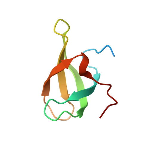
    Survival of motor neuron (SMN) functions in diverse biological pathways via recognition of symmetric dimethylarginine (Rme2s) on proteins by its Tudor domain, and deficiency of SMN leads to spinal muscular atrophy. Here we report a potent and selective antagonist with a 4-iminopyridine scaffold targeting the Tudor domain of SMN. Our structural and mutagenesis studies indicate that both the aromatic ring and imino groups of compound 1 contribute to its selective binding to SMN. Various on-target engagement assays support that compound 1 specifically recognizes SMN in a cellular context and prevents the interaction of SMN with the R1810me2s of RNA polymerase II subunit POLR2A, resulting in transcription termination and R-loop accumulation mimicking SMN depletion. Thus, in addition to the antisense, RNAi and CRISPR/Cas9 techniques, potent SMN antagonists could be used as an efficient tool to understand the biological functions of SMN.


  • Organizational Affiliation

    Jiangsu Key Laboratory of Neuropsychiatric Diseases and College of Pharmaceutical Sciences, Soochow University, Suzhou, Jiangsu, China. ylliu18@suda.edu.cn.


Macromolecules
Find similar proteins by:  (by identity cutoff)  |  3D Structure
Entity ID: 1
MoleculeChains Sequence LengthOrganismDetailsImage
Survival motor neuron protein67Homo sapiensMutation(s): 0 
Gene Names: SMN1SMNSMNTSMN2SMNC
UniProt & NIH Common Fund Data Resources
Find proteins for Q16637 (Homo sapiens)
Explore Q16637 
Go to UniProtKB:  Q16637
PHAROS:  Q16637
Entity Groups  
Sequence Clusters30% Identity50% Identity70% Identity90% Identity95% Identity100% Identity
UniProt GroupQ16637
Sequence Annotations
Expand
  • Reference Sequence
Experimental Data & Validation

Experimental Data

  • Method: X-RAY DIFFRACTION
  • Resolution: 1.75 Å
  • R-Value Free: 0.242 
  • R-Value Work: 0.165 
  • R-Value Observed: 0.173 
  • Space Group: P 65
  • Diffraction Data: https://doi.org/10.18430/m34qq6
Unit Cell:
Length ( Å )Angle ( ˚ )
a = 27.789α = 90
b = 27.789β = 90
c = 112.817γ = 120
Software Package:
Software NamePurpose
Aimlessdata scaling
REFMACrefinement
PDB_EXTRACTdata extraction
XDSdata reduction

Structure Validation

View Full Validation Report


Ligand Structure Quality Assessment 


Entry History 

Revision History  (Full details and data files)

  • Version 1.0: 2014-08-06
    Type: Initial release
  • Version 1.1: 2022-10-05
    Changes: Data collection, Database references, Derived calculations
  • Version 1.2: 2023-09-20
    Changes: Data collection, Refinement description