4PO7

Structure of the Sortilin:neurotensin complex at excess neurotensin concentration


Experimental Data Snapshot

  • Method: X-RAY DIFFRACTION
  • Resolution: 2.66 Å
  • R-Value Free: 0.212 
  • R-Value Work: 0.166 
  • R-Value Observed: 0.168 

wwPDB Validation   3D Report Full Report


Ligand Structure Quality Assessment 


This is version 3.1 of the entry. See complete history


Literature

Revisiting the structure of the Vps10 domain of human sortilin and its interaction with neurotensin.

Quistgaard, E.M.Grftehauge, M.K.Madsen, P.Pallesen, L.T.Christensen, B.Srensen, E.S.Nissen, P.Petersen, C.M.Thirup, S.S.

(2014) Protein Sci 23: 1291-1300

  • DOI: https://doi.org/10.1002/pro.2512
  • Primary Citation of Related Structures:  
    4PO7

  • PubMed Abstract: 

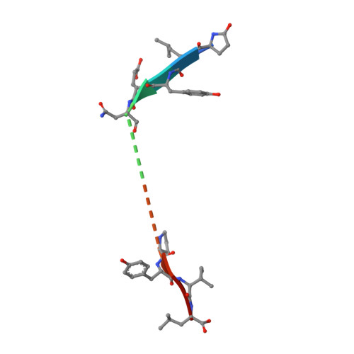
    Sortilin is a multifunctional receptor involved in sorting and apoptosis. We have previously reported a 2.0-Å structure of the Vps10 ectodomain in complex with one of its ligands, the tridecapeptide neurotensin. Here we set out to further characterize the structural properties of sortilin and its interaction with neurotensin. To this end, we have determined a new 2.7 Å structure using a crystal grown with a 10-fold increased concentration of neurotensin. Here a second peptide fragment was observed within the Vps10 β-propeller, which may in principle either represent a second molecule of neurotensin or the N-terminal part of the molecule bound at the previously identified binding site. However, in vitro binding experiments strongly favor the latter hypothesis. Neurotensin thus appears to bind with a 1:1 stoichiometry, and whereas the N-terminus does not bind on its own, it enhances the affinity in context of full-length neurotensin. We conclude that the N-terminus of neurotensin probably functions as an affinity enhancer for binding to sortilin by engaging the second binding site. Crystal packing differs partly from the previous structure, which may be due to variations in the degree and pattern of glycosylations. Consequently, a notable hydrophobic loop, not modeled previously, could now be traced. A computational analysis suggests that this and a neighboring loop may insert into the membrane and thus restrain movement of the Vps10 domain. We have, furthermore, mapped all N-linked glycosylations of CHO-expressed human sortilin by mass spectrometry and find that their locations are compatible with membrane insertion of the hydrophobic loops.


  • Organizational Affiliation

    Department of Molecular Biology and Genetics, MIND Centre, Aarhus University, Gustav Wieds Vej 10C, DK 8000, Aarhus C, Denmark; Department of Medical Biochemistry and Biophysics, Karolinska Institute, 17177, Stockholm, Sweden.


Macromolecules
Find similar proteins by:  (by identity cutoff)  |  3D Structure
Entity ID: 1
MoleculeChains Sequence LengthOrganismDetailsImage
Sortilin685Homo sapiensMutation(s): 1 
Gene Names: SORT1
UniProt & NIH Common Fund Data Resources
Find proteins for Q99523 (Homo sapiens)
Explore Q99523 
Go to UniProtKB:  Q99523
PHAROS:  Q99523
GTEx:  ENSG00000134243 
Entity Groups  
Sequence Clusters30% Identity50% Identity70% Identity90% Identity95% Identity100% Identity
UniProt GroupQ99523
Sequence Annotations
Expand
  • Reference Sequence

Find similar proteins by:  Sequence   |   3D Structure  

Entity ID: 2
MoleculeChains Sequence LengthOrganismDetailsImage
Neurotensin/neuromedin NB [auth N],
C [auth P]
13Homo sapiensMutation(s): 0 
UniProt & NIH Common Fund Data Resources
Find proteins for P30990 (Homo sapiens)
Explore P30990 
Go to UniProtKB:  P30990
PHAROS:  P30990
GTEx:  ENSG00000133636 
Entity Groups  
Sequence Clusters30% Identity50% Identity70% Identity90% Identity95% Identity100% Identity
UniProt GroupP30990
Sequence Annotations
Expand
  • Reference Sequence
Oligosaccharides

Help

Entity ID: 3
MoleculeChains Length2D Diagram Glycosylation3D Interactions
beta-D-mannopyranose-(1-4)-2-acetamido-2-deoxy-beta-D-glucopyranose-(1-4)-2-acetamido-2-deoxy-beta-D-glucopyranoseD [auth B],
E [auth C]
3N-Glycosylation
Glycosylation Resources
GlyTouCan:  G15407YE
GlyCosmos:  G15407YE
GlyGen:  G15407YE
Small Molecules
Ligands 2 Unique
IDChains Name / Formula / InChI Key2D Diagram3D Interactions
NAG
Query on NAG

Download Ideal Coordinates CCD File 
F [auth A]2-acetamido-2-deoxy-beta-D-glucopyranose
C8 H15 N O6
OVRNDRQMDRJTHS-FMDGEEDCSA-N
PG4
Query on PG4

Download Ideal Coordinates CCD File 
G [auth A]TETRAETHYLENE GLYCOL
C8 H18 O5
UWHCKJMYHZGTIT-UHFFFAOYSA-N
Modified Residues  1 Unique
IDChains TypeFormula2D DiagramParent
PCA
Query on PCA
B [auth N],
C [auth P]
L-PEPTIDE LINKINGC5 H7 N O3GLN
Experimental Data & Validation

Experimental Data

  • Method: X-RAY DIFFRACTION
  • Resolution: 2.66 Å
  • R-Value Free: 0.212 
  • R-Value Work: 0.166 
  • R-Value Observed: 0.168 
  • Space Group: C 1 2 1
Unit Cell:
Length ( Å )Angle ( ˚ )
a = 162.01α = 90
b = 78.65β = 126.62
c = 110.98γ = 90
Software Package:
Software NamePurpose
PHENIXrefinement

Structure Validation

View Full Validation Report


Ligand Structure Quality Assessment 


Entry History 

Deposition Data

Revision History  (Full details and data files)

  • Version 1.0: 2014-07-23
    Type: Initial release
  • Version 1.1: 2014-07-30
    Changes: Other
  • Version 1.2: 2014-09-24
    Changes: Database references
  • Version 1.3: 2018-03-07
    Changes: Advisory, Data collection
  • Version 2.0: 2019-12-25
    Changes: Database references, Derived calculations, Polymer sequence
  • Version 3.0: 2020-07-29
    Type: Remediation
    Reason: Carbohydrate remediation
    Changes: Atomic model, Data collection, Derived calculations, Structure summary
  • Version 3.1: 2020-10-21
    Changes: Data collection, Structure summary