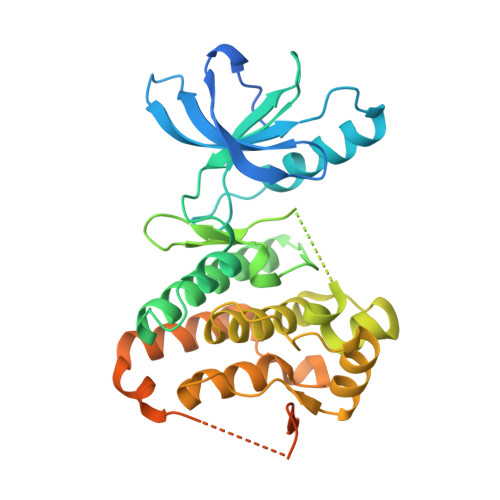
4P4C

Human EphA3 Kinase domain in complex with quinoxaline derivatives


Experimental Data Snapshot

  • Method: X-RAY DIFFRACTION
  • Resolution: 1.60 Å
  • R-Value Free: 0.202 
  • R-Value Work: 0.178 
  • R-Value Observed: 0.179 

wwPDB Validation   3D Report Full Report


Ligand Structure Quality Assessment 


This is version 1.3 of the entry. See complete history


Literature

Pyrrolo[3,2-b]quinoxaline Derivatives as Types I1/2 and II Eph Tyrosine Kinase Inhibitors: Structure-Based Design, Synthesis, and in Vivo Validation.

Unzue, A.Dong, J.Lafleur, K.Zhao, H.Frugier, E.Caflisch, A.Nevado, C.

(2014) J Med Chem 57: 6834-6844

  • DOI: https://doi.org/10.1021/jm5009242
  • Primary Citation of Related Structures:  
    4P4C, 4P5Q, 4P5Z

  • PubMed Abstract: 

    The X-ray crystal structures of the catalytic domain of the EphA3 tyrosine kinase in complex with two type I inhibitors previously discovered in silico (compounds A and B) were used to design type I1/2 and II inhibitors. Chemical synthesis of about 25 derivatives culminated in the discovery of compounds 11d (type I1/2), 7b, and 7g (both of type II), which have low-nanomolar affinity for Eph kinases in vitro and a good selectivity profile on a panel of 453 human kinases (395 nonmutant). Surface plasmon resonance measurements show a very slow unbinding rate (1/115 min) for inhibitor 7m. Slow dissociation is consistent with a type II binding mode in which the hydrophobic moiety (trifluoromethyl-benzene) of the inhibitor is deeply buried in a cavity originating from the displacement of the Phe side chain of the so-called DFG motif as observed in the crystal structure of compound 7m. The inhibitor 11d displayed good in vivo efficacy in a human breast cancer xenograft.


  • Organizational Affiliation

    Department of Chemistry and ‡Department of Biochemistry, University of Zürich , Winterthurerstrasse 190, CH-8057 Zürich, Switzerland.


Macromolecules
Find similar proteins by:  (by identity cutoff)  |  3D Structure
Entity ID: 1
MoleculeChains Sequence LengthOrganismDetailsImage
EPH receptor A3361Homo sapiensMutation(s): 0 
Gene Names: EPHA3
EC: 2.7.10.1
UniProt & NIH Common Fund Data Resources
Find proteins for P29320 (Homo sapiens)
Explore P29320 
Go to UniProtKB:  P29320
PHAROS:  P29320
GTEx:  ENSG00000044524 
Entity Groups  
Sequence Clusters30% Identity50% Identity70% Identity90% Identity95% Identity100% Identity
UniProt GroupP29320
Sequence Annotations
Expand
  • Reference Sequence
Small Molecules
Ligands 1 Unique
IDChains Name / Formula / InChI Key2D Diagram3D Interactions
25Q
Query on 25Q

Download Ideal Coordinates CCD File 
B [auth A]2-amino-1-(3-methoxyphenyl)-1H-pyrrolo[2,3-b]quinoxaline-3-carboxamide
C18 H15 N5 O2
CHHKMVNVTYIPGZ-UHFFFAOYSA-N
Experimental Data & Validation

Experimental Data

  • Method: X-RAY DIFFRACTION
  • Resolution: 1.60 Å
  • R-Value Free: 0.202 
  • R-Value Work: 0.178 
  • R-Value Observed: 0.179 
  • Space Group: P 1 21 1
Unit Cell:
Length ( Å )Angle ( ˚ )
a = 53.914α = 90
b = 38.164β = 101.59
c = 75.527γ = 90
Software Package:
Software NamePurpose
SCALAdata scaling
XDSdata scaling
PDB_EXTRACTdata extraction
PHASERphasing
PHENIXrefinement

Structure Validation

View Full Validation Report


Ligand Structure Quality Assessment 


Entry History & Funding Information

Deposition Data


Funding OrganizationLocationGrant Number
KrebsligaSwitzerland--
Swiss National Science FoundationSwitzerland--

Revision History  (Full details and data files)

  • Version 1.0: 2014-08-13
    Type: Initial release
  • Version 1.1: 2014-10-01
    Changes: Database references
  • Version 1.2: 2017-11-22
    Changes: Database references, Derived calculations, Other, Refinement description, Source and taxonomy
  • Version 1.3: 2023-09-27
    Changes: Data collection, Database references, Refinement description