4OYK

Structure of HOIP PUB domain bound to OTULIN PIM


Experimental Data Snapshot

  • Method: X-RAY DIFFRACTION
  • Resolution: 2.00 Å
  • R-Value Free: 0.237 
  • R-Value Work: 0.201 
  • R-Value Observed: 0.202 

wwPDB Validation   3D Report Full Report


This is version 1.1 of the entry. See complete history


Literature

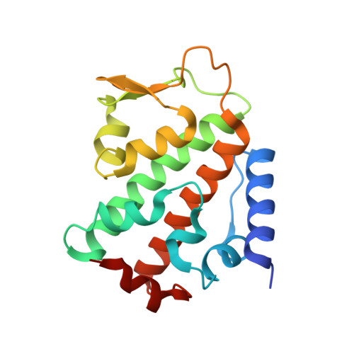
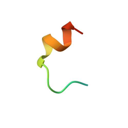
Molecular Basis and Regulation of OTULIN-LUBAC Interaction.

Elliott, P.R.Nielsen, S.V.Marco-Casanova, P.Fiil, B.K.Keusekotten, K.Mailand, N.Freund, S.M.Gyrd-Hansen, M.Komander, D.

(2014) Mol Cell 54: 335-348

  • DOI: https://doi.org/10.1016/j.molcel.2014.03.018
  • Primary Citation of Related Structures:  
    4OYJ, 4OYK

  • PubMed Abstract: 

    The linear ubiquitin (Ub) chain assembly complex (LUBAC) generates Met1-linked "linear" Ub chains that regulate the activation of the nuclear factor κB (NFκB) transcription factor and other processes. We recently discovered OTULIN as a deubiquitinase that specifically cleaves Met1-linked polyUb. Now, we show that OTULIN binds via a conserved PUB-interacting motif (PIM) to the PUB domain of the LUBAC component HOIP. Crystal structures and nuclear magnetic resonance experiments reveal the molecular basis for the high-affinity interaction and explain why OTULIN binds the HOIP PUB domain specifically. Analysis of LUBAC-induced NFκB signaling suggests that OTULIN needs to be present on LUBAC in order to restrict Met1-polyUb signaling. Moreover, LUBAC-OTULIN complex formation is regulated by OTULIN phosphorylation in the PIM. Phosphorylation of OTULIN prevents HOIP binding, whereas unphosphorylated OTULIN is part of the endogenous LUBAC complex. Our work exemplifies how coordination of ubiquitin assembly and disassembly activities in protein complexes regulates individual Ub linkage types.


  • Organizational Affiliation

    Medical Research Council Laboratory of Molecular Biology, Francis Crick Avenue, Cambridge, CB2 0QH, UK.


Macromolecules
Find similar proteins by:  (by identity cutoff)  |  3D Structure
Entity ID: 1
MoleculeChains Sequence LengthOrganismDetailsImage
E3 ubiquitin-protein ligase RNF31
A, B
177Homo sapiensMutation(s): 0 
Gene Names: RNF31ZIBRA
EC: 6.3.2
UniProt & NIH Common Fund Data Resources
Find proteins for Q96EP0 (Homo sapiens)
Explore Q96EP0 
Go to UniProtKB:  Q96EP0
PHAROS:  Q96EP0
GTEx:  ENSG00000092098 
Entity Groups  
Sequence Clusters30% Identity50% Identity70% Identity90% Identity95% Identity100% Identity
UniProt GroupQ96EP0
Sequence Annotations
Expand
  • Reference Sequence

Find similar proteins by:  Sequence   |   3D Structure  

Entity ID: 2
MoleculeChains Sequence LengthOrganismDetailsImage
Ubiquitin thioesterase otulin
C, D
19Homo sapiensMutation(s): 0 
EC: 3.4.19.12
UniProt & NIH Common Fund Data Resources
Find proteins for Q96BN8 (Homo sapiens)
Explore Q96BN8 
Go to UniProtKB:  Q96BN8
PHAROS:  Q96BN8
GTEx:  ENSG00000154124 
Entity Groups  
Sequence Clusters30% Identity50% Identity70% Identity90% Identity95% Identity100% Identity
UniProt GroupQ96BN8
Sequence Annotations
Expand
  • Reference Sequence
Experimental Data & Validation

Experimental Data

  • Method: X-RAY DIFFRACTION
  • Resolution: 2.00 Å
  • R-Value Free: 0.237 
  • R-Value Work: 0.201 
  • R-Value Observed: 0.202 
  • Space Group: P 61
Unit Cell:
Length ( Å )Angle ( ˚ )
a = 64.049α = 90
b = 64.049β = 90
c = 172.022γ = 120
Software Package:
Software NamePurpose
PHENIXrefinement

Structure Validation

View Full Validation Report


Entry History 

Deposition Data

Revision History  (Full details and data files)

  • Version 1.0: 2014-05-21
    Type: Initial release
  • Version 1.1: 2023-09-27
    Changes: Data collection, Database references, Derived calculations, Other, Refinement description, Source and taxonomy