4OEW

Crystal structure of the PDE5A1 catalytic domain in complex with novel inhibitors


Experimental Data Snapshot

  • Method: X-RAY DIFFRACTION
  • Resolution: 2.44 Å
  • R-Value Free: 0.259 
  • R-Value Work: 0.206 
  • R-Value Observed: 0.208 

wwPDB Validation   3D Report Full Report


Ligand Structure Quality Assessment 


This is version 1.1 of the entry. See complete history


Literature

Thermodynamic and structural characterization of halogen bonding in protein-ligand interactions: a case study of PDE5 and its inhibitors.

Ren, J.He, Y.Chen, W.Chen, T.Wang, G.Wang, Z.Xu, Z.Luo, X.Zhu, W.Jiang, H.Shen, J.Xu, Y.

(2014) J Med Chem 57: 3588-3593

  • DOI: https://doi.org/10.1021/jm5002315
  • Primary Citation of Related Structures:  
    4OEW, 4OEX

  • PubMed Abstract: 

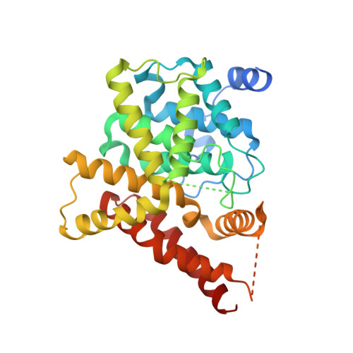
    The significance of halogen bonding in protein-ligand interactions has been recognized recently. We present here the first comprehensive thermodynamic and structural characterization of halogen bonding in PDE5-inhibitor interactions. ITC studies reveal that binding strength of the halogen bonding between chlorine, bromine, and iodine of inhibitor and the protein is -1.57, -3.09, and -5.59 kJ/mol, respectively. The halogens interact with the designed residue Y612 and an unexpected buried water molecule.


  • Organizational Affiliation

    CAS Key Laboratory of Receptor Research, Drug Discovery and Design Center, Shanghai Institute of Materia Medica, Chinese Academy of Sciences (CAS) , 555 Zuchongzhi Road, Shanghai 201203, China.


Macromolecules
Find similar proteins by:  (by identity cutoff)  |  3D Structure
Entity ID: 1
MoleculeChains Sequence LengthOrganismDetailsImage
cGMP-specific 3',5'-cyclic phosphodiesterase347Homo sapiensMutation(s): 0 
Gene Names: PDE5PDE5A
EC: 3.1.4.35
UniProt & NIH Common Fund Data Resources
Find proteins for O76074 (Homo sapiens)
Explore O76074 
Go to UniProtKB:  O76074
PHAROS:  O76074
GTEx:  ENSG00000138735 
Entity Groups  
Sequence Clusters30% Identity50% Identity70% Identity90% Identity95% Identity100% Identity
UniProt GroupO76074
Sequence Annotations
Expand
  • Reference Sequence
Small Molecules
Ligands 3 Unique
IDChains Name / Formula / InChI Key2D Diagram3D Interactions
5IO
Query on 5IO

Download Ideal Coordinates CCD File 
D [auth A]6-ethyl-5-iodo-2-{5-[(4-methylpiperazin-1-yl)sulfonyl]-2-propoxyphenyl}pyrimidin-4(3H)-one
C20 H27 I N4 O4 S
CLYYLXSJJMHCHN-UHFFFAOYSA-N
ZN
Query on ZN

Download Ideal Coordinates CCD File 
B [auth A]ZINC ION
Zn
PTFCDOFLOPIGGS-UHFFFAOYSA-N
MG
Query on MG

Download Ideal Coordinates CCD File 
C [auth A]MAGNESIUM ION
Mg
JLVVSXFLKOJNIY-UHFFFAOYSA-N
Binding Affinity Annotations 
IDSourceBinding Affinity
5IO BindingDB:  4OEW Kd: 150 (nM) from 1 assay(s)
Binding MOAD:  4OEW Kd: 64 (nM) from 1 assay(s)
Experimental Data & Validation

Experimental Data

  • Method: X-RAY DIFFRACTION
  • Resolution: 2.44 Å
  • R-Value Free: 0.259 
  • R-Value Work: 0.206 
  • R-Value Observed: 0.208 
  • Space Group: P 31 2 1
Unit Cell:
Length ( Å )Angle ( ˚ )
a = 74.12α = 90
b = 74.12β = 90
c = 131.8γ = 120
Software Package:
Software NamePurpose
XSCALEdata scaling
PHASERphasing
PHENIXrefinement
PDB_EXTRACTdata extraction
Blu-Icedata collection
XDSdata reduction

Structure Validation

View Full Validation Report


Ligand Structure Quality Assessment 


Entry History 

Deposition Data

Revision History  (Full details and data files)

  • Version 1.0: 2015-04-01
    Type: Initial release
  • Version 1.1: 2024-02-28
    Changes: Data collection, Database references, Derived calculations