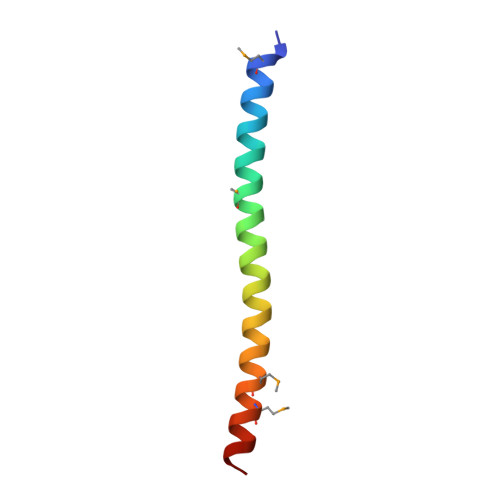
Striatins contain a noncanonical coiled coil that binds protein phosphatase 2A A subunit to form a 2:2 heterotetrameric core of striatin-interacting phosphatase and kinase (STRIPAK) complex.
Chen, C., Shi, Z., Zhang, W., Chen, M., He, F., Zhang, Z., Wang, Y., Feng, M., Wang, W., Zhao, Y., Brown, J.H., Jiao, S., Zhou, Z.(2014) J Biol Chem 289: 9651-9661
- PubMed: 24550388
- DOI: https://doi.org/10.1074/jbc.M113.529297
- Primary Citation of Related Structures:
4N6J - PubMed Abstract:
The protein phosphatase 2A (PP2A) and kinases such as germinal center kinase III (GCKIII) can interact with striatins to form a supramolecular complex called striatin-interacting phosphatase and kinase (STRIPAK) complex. Despite the fact that the STRIPAK complex regulates multiple cellular events, it remains only partially understood how this complex itself is assembled and regulated for differential biological functions. Our recent work revealed the activation mechanism of GCKIIIs by MO25, as well as how GCKIIIs heterodimerize with CCM3, a molecular bridge between GCKIII and striatins. Here we dissect the structural features of the coiled coil domain of striatin 3, a novel type of PP2A regulatory subunit that functions as a scaffold for the assembly of the STRIPAK complex. We have determined the crystal structure of a selenomethionine-labeled striatin 3 coiled coil domain, which shows it to assume a parallel dimeric but asymmetric conformation containing a large bend. This result combined with a number of biophysical analyses provide evidence that the coiled coil domain of striatin 3 and the PP2A A subunit form a stable core complex with a 2:2 stoichiometry. Structure-based mutational studies reveal that homodimerization of striatin 3 is essential for its interaction with PP2A and therefore assembly of the STRIPAK complex. Wild-type striatin 3 but not the mutants defective in PP2A binding strongly suppresses apoptosis of Jurkat cells induced by the GCKIII kinase MST3, most likely through a mechanism in which striatin recruits PP2A to negatively regulate the activation of MST3. Collectively, our work provides structural insights into the organization of the STRIPAK complex and will facilitate further functional studies.
Organizational Affiliation:
From the National Center for Protein Science Shanghai, State Key Laboratory of Cell Biology, Institute of Biochemistry and Cell Biology, Shanghai Institutes for Biological Sciences, Chinese Academy of Sciences, Shanghai 200031, China.