DJ-1 Is a Copper Chaperone Acting on SOD1 Activation.
Girotto, S., Cendron, L., Bisaglia, M., Tessari, I., Mammi, S., Zanotti, G., Bubacco, L.(2014) J Biol Chem 289: 10887-10899
- PubMed: 24567322
- DOI: https://doi.org/10.1074/jbc.M113.535112
- Primary Citation of Related Structures:
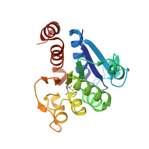
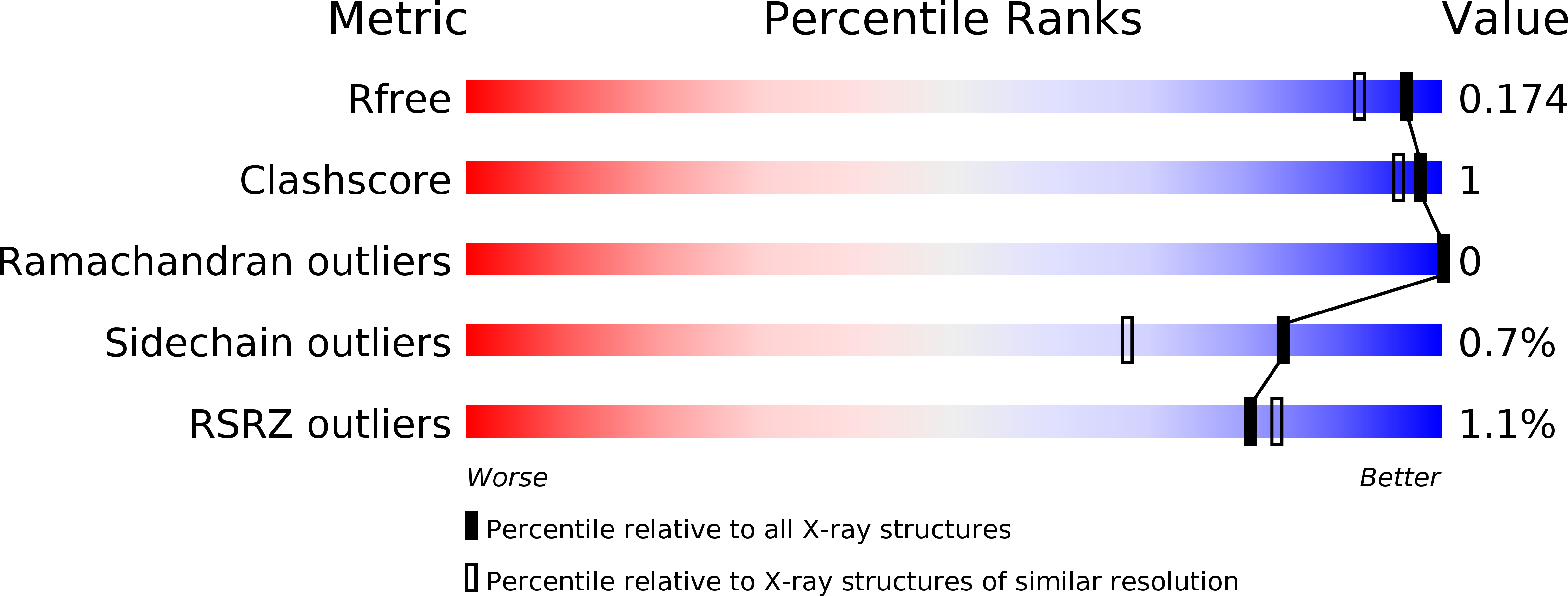
4MNT, 4MTC, 4N0M, 4N12 - PubMed Abstract:
Lack of oxidative stress control is a common and often prime feature observed in many neurodegenerative diseases. Both DJ-1 and SOD1, proteins involved in familial Parkinson disease and amyotrophic lateral sclerosis, respectively, play a protective role against oxidative stress. Impaired activity and modified expression of both proteins have been observed in different neurodegenerative diseases. A potential cooperative action of DJ-1 and SOD1 in the same oxidative stress response pathway may be suggested based on a copper-mediated interaction between the two proteins reported here. To investigate the mechanisms underlying the antioxidative function of DJ-1 in relation to SOD1 activity, we investigated the ability of DJ-1 to bind copper ions. We structurally characterized a novel copper binding site involving Cys-106, and we investigated, using different techniques, the kinetics of DJ-1 binding to copper ions. The copper transfer between the two proteins was also examined using both fluorescence spectroscopy and specific biochemical assays for SOD1 activity. The structural and functional analysis of the novel DJ-1 copper binding site led us to identify a putative role for DJ-1 as a copper chaperone. Alteration of the coordination geometry of the copper ion in DJ-1 may be correlated to the physiological role of the protein, to a potential failure in metal transfer to SOD1, and to successive implications in neurodegenerative etiopathogenesis.
Organizational Affiliation:
Department of Chemical Sciences, University of Padova, via Marzolo, 1 35131 Padova, Italy; Department of Drug Discovery and Development, Istituto Italiano di Tecnologia, Via Morego, 30 16163 Genoa, Italy.