4MYW

Structure of HSV-2 gD bound to nectin-1


Experimental Data Snapshot

  • Method: X-RAY DIFFRACTION
  • Resolution: 3.19 Å
  • R-Value Free: 0.247 
  • R-Value Work: 0.196 
  • R-Value Observed: 0.198 

wwPDB Validation   3D Report Full Report


Ligand Structure Quality Assessment 


This is version 2.2 of the entry. See complete history


Literature

Crystal structure of herpes simplex virus 2 gD bound to nectin-1 reveals a conserved mode of receptor recognition.

Lu, G.Zhang, N.Qi, J.Li, Y.Chen, Z.Zheng, C.Gao, G.F.Yan, J.

(2014) J Virol 88: 13678-13688

  • DOI: https://doi.org/10.1128/JVI.01906-14
  • Primary Citation of Related Structures:  
    4MYV, 4MYW

  • PubMed Abstract: 

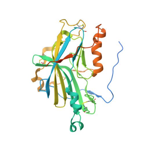
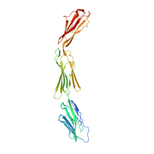
    Herpes simplex virus 1 (HSV-1) and HSV-2 are among the most prevalent human pathogens. Both viruses can recognize, via the surface envelope glycoprotein D (gD), human nectin-1 as a functional receptor. Previous studies have successfully elucidated the molecular basis of the binding between HSV-1 gD and nectin-1 by cocrystallography. Despite a high sequence identity between HSV-1 and HSV-2 gDs, the atomic intermolecule details for the HSV-2-gD/nectin-1 interaction remain elusive. Here, we report the crystal structures of both the unbound and the nectin-1-bound HSV-2 gDs. The free-gD structure expectedly comprises an IgV-like core and the surface-exposed terminal extensions as observed in its HSV-1 counterpart but lacks traceable electron densities for a large portion of the terminal elements. These terminal residues were clearly traced in the complex structure as a definitive loop in the N terminus and an α-helix in the C terminus, thereby showing a conserved nectin-1-binding mode as reported for HSV-1 gD. The interface residues in nectin-1 were further mutated and tested for the gD interaction by surface plasmon resonance. The resultant binding patterns were similar for HSV-1 and HSV-2 gDs, further supporting a homologous receptor-binding basis by the two viruses for nectin-1. These data, together with a cell-based fusion assay showing a cross-inhibition of the gD/nectin-1-mediated cell-cell fusion by soluble HSV-1 and HSV-2 gDs, provided solid structural and functional evidence that HSV-1 and HSV-2 recognize nectin-1 via the same binding mode. Finally, we also demonstrated that nectin-1 I80 is an important residue involved in gD interaction.


  • Organizational Affiliation

    CAS Key Laboratory of Pathogenic Microbiology and Immunology, Institute of Microbiology, Chinese Academy of Sciences, Beijing, China.


Macromolecules
Find similar proteins by:  (by identity cutoff)  |  3D Structure
Entity ID: 1
MoleculeChains Sequence LengthOrganismDetailsImage
Envelope glycoprotein D
A, C
294Human herpesvirus 2 strain 333Mutation(s): 0 
Gene Names: gDUS6
UniProt
Find proteins for P03172 (Human herpesvirus 2 (strain 333))
Explore P03172 
Go to UniProtKB:  P03172
Entity Groups  
Sequence Clusters30% Identity50% Identity70% Identity90% Identity95% Identity100% Identity
UniProt GroupP03172
Sequence Annotations
Expand
  • Reference Sequence
Find similar proteins by:  (by identity cutoff)  |  3D Structure
Entity ID: 2
MoleculeChains Sequence LengthOrganismDetailsImage
Poliovirus receptor-related protein 1
B, D
331Homo sapiensMutation(s): 0 
Gene Names: HVECnectin-1PRR1PVRL1
UniProt & NIH Common Fund Data Resources
Find proteins for Q15223 (Homo sapiens)
Explore Q15223 
Go to UniProtKB:  Q15223
PHAROS:  Q15223
GTEx:  ENSG00000110400 
Entity Groups  
Sequence Clusters30% Identity50% Identity70% Identity90% Identity95% Identity100% Identity
UniProt GroupQ15223
Sequence Annotations
Expand
  • Reference Sequence
Oligosaccharides

Help

Entity ID: 3
MoleculeChains Length2D Diagram Glycosylation3D Interactions
2-acetamido-2-deoxy-beta-D-glucopyranose-(1-4)-2-acetamido-2-deoxy-beta-D-glucopyranose
E, F
2N-Glycosylation
Glycosylation Resources
GlyTouCan:  G42666HT
GlyCosmos:  G42666HT
GlyGen:  G42666HT
Small Molecules
Ligands 1 Unique
IDChains Name / Formula / InChI Key2D Diagram3D Interactions
NAG
Query on NAG

Download Ideal Coordinates CCD File 
G [auth A],
H [auth C]
2-acetamido-2-deoxy-beta-D-glucopyranose
C8 H15 N O6
OVRNDRQMDRJTHS-FMDGEEDCSA-N
Experimental Data & Validation

Experimental Data

  • Method: X-RAY DIFFRACTION
  • Resolution: 3.19 Å
  • R-Value Free: 0.247 
  • R-Value Work: 0.196 
  • R-Value Observed: 0.198 
  • Space Group: P 2 21 21
Unit Cell:
Length ( Å )Angle ( ˚ )
a = 53.337α = 90
b = 170.857β = 90
c = 192.251γ = 90
Software Package:
Software NamePurpose
Blu-Icedata collection
AMoREphasing
PHENIXrefinement
HKL-2000data reduction
HKL-2000data scaling

Structure Validation

View Full Validation Report


Ligand Structure Quality Assessment 


Entry History 

Deposition Data

Revision History  (Full details and data files)

  • Version 1.0: 2014-10-01
    Type: Initial release
  • Version 2.0: 2020-07-29
    Type: Remediation
    Reason: Carbohydrate remediation
    Changes: Advisory, Atomic model, Data collection, Database references, Derived calculations, Structure summary
  • Version 2.1: 2022-08-24
    Changes: Database references, Structure summary
  • Version 2.2: 2023-11-08
    Changes: Data collection, Refinement description