4MNV

Crystal structure of bicyclic peptide UK729 bound as an acyl-enzyme intermediate to urokinase-type plasminogen activator (uPA)


Experimental Data Snapshot

  • Method: X-RAY DIFFRACTION
  • Resolution: 1.80 Å
  • R-Value Free: 0.216 
  • R-Value Work: 0.189 
  • R-Value Observed: 0.190 

wwPDB Validation   3D Report Full Report


Ligand Structure Quality Assessment 


This is version 1.2 of the entry. See complete history


Literature

Peptide ligands stabilized by small molecules.

Chen, S.Bertoldo, D.Angelini, A.Pojer, F.Heinis, C.

(2014) Angew Chem Int Ed Engl 53: 1602-1606

  • DOI: https://doi.org/10.1002/anie.201309459
  • Primary Citation of Related Structures:  
    4MNV, 4MNW, 4MNX, 4MNY

  • PubMed Abstract: 

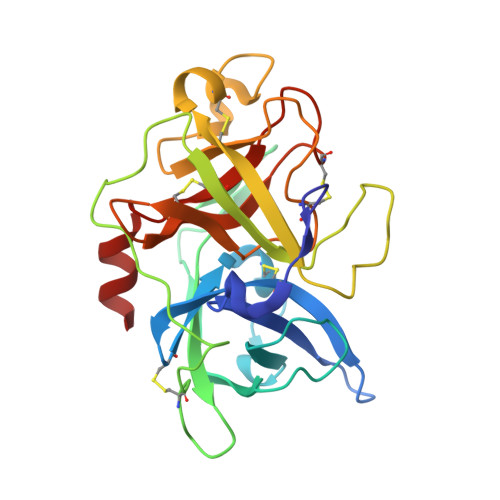
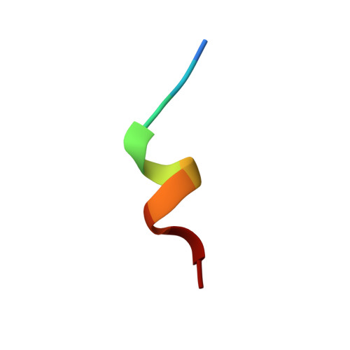
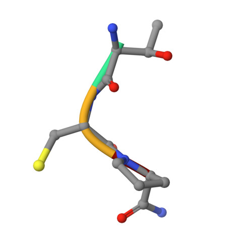
    Bicyclic peptides generated through directed evolution by using phage display offer an attractive ligand format for the development of therapeutics. Being nearly 100-fold smaller than antibodies, they promise advantages such as access to chemical synthesis, efficient diffusion into tissues, and needle-free application. However, unlike antibodies, they do not have a folded structure in solution and thus bind less well. We developed bicyclic peptides with hydrophilic chemical structures at their center to promote noncovalent intramolecular interactions, thereby stabilizing the peptide conformation. The sequences of the peptides isolated by phage display from large combinatorial libraries were strongly influenced by the type of small molecule used in the screen, thus suggesting that the peptides fold around the small molecules. X-ray structure analysis revealed that the small molecules indeed formed hydrogen bonds with the peptides. These noncovalent interactions stabilize the peptide-protein complexes and contribute to the high binding affinity.


  • Organizational Affiliation

    Institute of Chemical Sciences and Engineering, Ecole Polytechnique Fédérale de Lausanne, 1015 Lausanne (Switzerland).


Macromolecules
Find similar proteins by:  (by identity cutoff)  |  3D Structure
Entity ID: 1
MoleculeChains Sequence LengthOrganismDetailsImage
Urokinase-type plasminogen activator chain B245Homo sapiensMutation(s): 2 
Gene Names: PLAU
EC: 3.4.21.73
UniProt & NIH Common Fund Data Resources
Find proteins for P00749 (Homo sapiens)
Explore P00749 
Go to UniProtKB:  P00749
PHAROS:  P00749
GTEx:  ENSG00000122861 
Entity Groups  
Sequence Clusters30% Identity50% Identity70% Identity90% Identity95% Identity100% Identity
UniProt GroupP00749
Sequence Annotations
Expand
  • Reference Sequence

Find similar proteins by:  Sequence   |   3D Structure  

Entity ID: 2
MoleculeChains Sequence LengthOrganismDetailsImage
acyl-enzyme intermediate of bicyclic peptide UK72910N/AMutation(s): 0 
Entity Groups  
Sequence Clusters30% Identity50% Identity70% Identity90% Identity95% Identity100% Identity
Sequence Annotations
Expand
  • Reference Sequence

Find similar proteins by:  Sequence   |   3D Structure  

Entity ID: 3
MoleculeChains Sequence LengthOrganismDetailsImage
acyl-enzyme intermediate of bicyclic peptide UK7294N/AMutation(s): 0 
Sequence Annotations
Expand
  • Reference Sequence
Experimental Data & Validation

Experimental Data

  • Method: X-RAY DIFFRACTION
  • Resolution: 1.80 Å
  • R-Value Free: 0.216 
  • R-Value Work: 0.189 
  • R-Value Observed: 0.190 
  • Space Group: P 21 21 21
Unit Cell:
Length ( Å )Angle ( ˚ )
a = 52.31α = 90
b = 54.06β = 90
c = 79.67γ = 90
Software Package:
Software NamePurpose
PHASERphasing
REFMACrefinement
MOSFLMdata reduction
SCALAdata scaling

Structure Validation

View Full Validation Report


Ligand Structure Quality Assessment 


Entry History 

Deposition Data

Revision History  (Full details and data files)

  • Version 1.0: 2014-02-05
    Type: Initial release
  • Version 1.1: 2014-02-26
    Changes: Database references
  • Version 1.2: 2017-11-15
    Changes: Refinement description